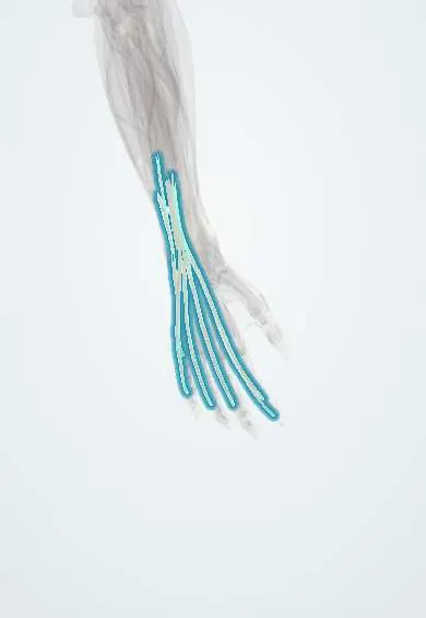
Extensor tendons !
The finger extensor tendons run over the back of the hand to the fingers; they straighten the fingers at the knuckles.
Official name: Extensor digitorum tendons
Opens in the Durgical app, no sign-up needed.
What it is
Four tendons across the back of the hand to the fingers, linked by small bands.
What it does
Straighten the fingers at the knuckles.
Key facts
- Attaches
- Four tendons in the 4th dorsal compartment (with the extensor indicis) to the extensor expansions of fingers 2–5; linked on the back of the hand by juncturae tendinum
- Function
- Extend the MCP joints, and the interphalangeal joints through the expansion; the juncturae can hide a single cut tendon
- Tight in
- Finger and wrist flexion
Relations
- Course
- From the extensor digitorum belly the four tendons pass under the extensor retinaculum in the 4th dorsal compartment, fan out over the back of the hand and widen into the extensor hood over each knuckle
- Deep
- Extensor indicis tendon in the same compartment, with the end of the posterior interosseous nerve on its floor; on the hand, the metacarpals and dorsal interossei
- Superficial
- Extensor retinaculum at the wrist, then the dorsal venous network of the hand and the dorsal branches of the superficial radial and ulnar nerves
- Lateral
- EPL tendon in the 3rd compartment, crossing obliquely toward the thumb
- Medial
- Extensor digiti minimi in the 5th compartment, over the distal radioulnar joint
How to find it
With the fingers extended against resistance, the four tendons stand out on the back of the hand; the linking bands can be seen as oblique cross-bands near the knuckles. Each tendon should stay centred over its knuckle as the fist closes.
Examination
- MCP extension against resistance, one finger at a time: weakness or a lag points to a cut tendon; a linking band may keep some extension, so strength matters more than movement
- Tenodesis effect: wrist flexion normally extends the MCPs; if it does in a person who cannot extend actively, the tendons are intact and a posterior interosseous nerve palsy is more likely
On imaging
Ultrasound is the first choice: in real time it shows tendon continuity, the gap of a cut and sagittal band tears, with the tendon slipping off the knuckle as the fist closes. MRI shows the same on axial images. Radiographs look for a fragment or a tooth in a fight bite over the MCP joint and for a bony mallet fracture at the DIP.
Clinical notes
- Rheumatoid arthritis: attrition rupture over a prominent ulnar head (Vaughan-Jackson syndrome) starts with the little finger and moves radially
- Sagittal band rupture, usually the radial band of the middle finger ('boxer's knuckle'), makes the tendon snap ulnarly as the fist closes
- Zone V cuts from a fight bite carry bacteria into the MCP joint and are explored and washed out
Anatomical variants
- The tendon to the little finger is often thin, absent or replaced by a linking band from the ring finger tendon
- Duplicated slips are common, and the extensor indicis origin and slips vary
Related conditions
- Rheumatoid arthritis
- Mallet finger
- Boxer fracture (fifth metacarpal neck)
- Rheumatoid arthritis of the hand and wrist
Test yourself
Think of the answer, then tap the question to see it.
In which compartment do the extensor digitorum tendons run, and what links them on the back of the hand?
The 4th dorsal compartment, with the extensor indicis; they are linked by the juncturae tendinum.
Why is a tooth wound over a knuckle (extensor zone V) explored?
A fight bite is assumed to have entered the MCP joint, carrying bacteria in; it is explored and washed out.
Why can the index and little fingers extend independently?
Each has a second extensor (extensor indicis, extensor digiti minimi) on the ulnar side of its extensor digitorum tendon.
What happens to an extensor tendon when its sagittal band tears?
It slips off the knuckle into the groove, usually the middle finger snapping ulnarly as the fist closes.
Educational information, not medical advice. For any symptom, see a health professional.
Its names
- Official Latin (TA2)
- tendines musculi extensoris digitorum TA2 2500
- Official Spanish (DeCS)
- Tendones del músculo extensor de los dedos
- English (TA2)
- Extensor digitorum tendons
Related
- Capitate
- Hamate
- Lunate
- 2nd metacarpal
- 3rd metacarpal
- 4th metacarpal
- 5th metacarpal
- Middle phalanx, index finger
Sources: Terminologia Anatomica (TA2); Anatomy, Shoulder and Upper Limb, Hand Compartments; Anatomy, Shoulder and Upper Limb, Wrist Extensor Muscles.
Spotted a mistake?We are always improving for the community. Tell us what is wrong and our team will review it.Report a mistake
Representative model, for educational purposes only.