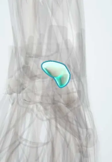
Scaphoid !
The scaphoid is the wrist bone at the base of the thumb; it is the carpal bone most often broken, and its poor blood supply slows healing.
Opens in the Durgical app, no sign-up needed.
What it is
The boat-shaped wrist bone at the base of the thumb; it links the two rows of wrist bones.
What it does
One of the eight carpal bones that let the wrist bend and carry load to the forearm.
Key facts
- Joints with
- Radius, lunate, capitate, trapezium, trapezoid
- Landmarks
- Tubercle, waist (common fracture site); floor of the anatomical snuffbox
Relations
- Above
- Scaphoid fossa of the distal radius; the radial styloid lies alongside its waist
- Below
- Trapezium and trapezoid at the STT joint
- Medial
- Lunate, joined by the scapholunate ligament; capitate distally
- Lateral
- Floor of the anatomical snuffbox: the radial artery crosses it between the EPL tendon behind and the APL and EPB tendons in front
- In front
- Tubercle with the flexor retinaculum and the abductor pollicis brevis; the flexor carpi radialis tendon passes close by on its way to the trapezium
- Behind
- Dorsal ridge where branches of the radial artery enter; dorsal radiocarpal ligament
How to find it
With the thumb extended, the anatomical snuffbox appears between the EPL tendon and the APL and EPB tendons; the waist of the scaphoid is under its floor and is felt best with the wrist in ulnar deviation. The tubercle is felt on the palm at the base of the thenar eminence, near the distal wrist crease, and stands out with the wrist extended.
Examination
- Snuffbox tenderness: sensitive for fracture but not specific
- Scaphoid tubercle tenderness and pain on axial compression of the thumb add to the suspicion of fracture
- Watson scaphoid shift test: the examiner presses on the tubercle while moving the wrist from ulnar to radial deviation; pain or a clunk on release suggests scapholunate instability
On imaging
PA, lateral, oblique and a PA view in ulnar deviation (scaphoid view), which lays the bone out flat. If X-rays are normal but suspicion remains, MRI (or CT) or repeat films. On the PA view a scapholunate gap over about 3 mm (Terry Thomas sign) and a ring-shaped scaphoid suggest ligament tear; on the lateral, a scapholunate angle over about 60° suggests DISI.
Common problems
- Fracture from a fall on an outstretched hand; pain in the 'snuffbox' at the thumb side of the wrist.
- Poor blood supply: it may heal slowly, not heal (nonunion) or lose its blood supply.
- Long-term, an unhealed scaphoid leads to wrist arthritis.
How it is diagnosed
Tenderness in the snuffbox. Special scaphoid X-rays; a fracture can be invisible at first, so a cast and repeat X-rays, or an MRI or CT, are used.
Treatment
- Cast for undisplaced fractures (often several weeks).
- A screw across the fracture for displaced fractures, or to return to activity sooner.
- Bone graft and fixation for fractures that don't heal.
Clinical notes
- Long-standing nonunion leads to scaphoid nonunion advanced collapse (SNAC wrist), a predictable pattern of arthritis
- A dense, white proximal pole after a fracture suggests avascular necrosis; necrosis without a fracture is Preiser disease
- Distal pole fractures heal well, waist fractures more slowly and proximal pole fractures worst
- In a trans-scaphoid perilunate fracture-dislocation the fracture line runs through the scaphoid instead of the scapholunate ligament
Anatomical variants
- Bipartite scaphoid, which is rare and hard to tell apart from an old nonunion
- Os centrale, a small accessory bone between the scaphoid, trapezoid and capitate
Development
Ossifies around age 4–6. In children, fractures more often involve the distal third and usually heal well.
Memory aid
Carpal bones from lateral to medial, proximal row then distal row: "Some Lovers Try Positions That They Can't Handle" (scaphoid, lunate, triquetrum, pisiform; trapezium, trapezoid, capitate, hamate).
Related conditions
Test yourself
Think of the answer, then tap the question to see it.
Why do waist and proximal pole fractures of the scaphoid risk nonunion and avascular necrosis?
Blood enters mainly through the dorsal ridge and flows from distal to proximal, so the proximal pole loses its supply.
Which tendons bound the anatomical snuffbox, whose floor is formed by the scaphoid?
The extensor pollicis longus behind and the abductor pollicis longus and extensor pollicis brevis in front; the radial artery crosses the floor.
If first X-rays of a suspected scaphoid fracture are normal, what is the next step?
MRI (or CT) or repeat films, because a scaphoid fracture may not show on the first X-rays.
What do the Terry Thomas sign and a scapholunate angle over about 60° suggest?
A scapholunate ligament tear: a gap over about 3 mm on the PA view, and DISI on the lateral view.
What does the Watson scaphoid shift test assess?
Scapholunate instability: pressure on the scaphoid tubercle while the wrist moves from ulnar to radial deviation gives pain or a clunk on release.
Educational information, not medical advice. For any symptom, see a health professional.
Its names
- Official Latin (TA2)
- os scaphoideum TA2 1250
- Official Spanish (DeCS)
- Hueso escafoides
- English (TA2)
- Scaphoid
- Also called
- navicular (of the hand)
Related
- Radial artery
- APB
- Wrist cartilage (radius)
- Dorsal intercarpal ligament
- Dorsal radiocarpal ligament
- Midcarpal ligaments
- Palmar wrist ligaments
- Scapholunate ligament
Sources: Terminologia Anatomica (TA2); Anatomy, Shoulder and Upper Limb, Hand Carpal Bones.
Spotted a mistake?We are always improving for the community. Tell us what is wrong and our team will review it.Report a mistake
Representative model, for educational purposes only.