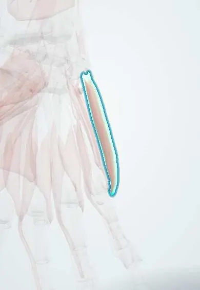
Abductor digiti minimi !
The abductor digiti minimi is the outermost muscle of the little-finger mound of the palm; it spreads the little finger away.
Opens in the Durgical app, no sign-up needed.
What it is
The muscle along the little-finger edge of the palm (hypothenar eminence).
What it does
Spreads the little finger away.
Key facts
- Origin
- Pisiform and FCU tendon
- Insertion
- Medial base of the proximal phalanx of the little finger
- Action
- Abducts the little finger
- Nerve
- Deep branch of the ulnar nerve (C8–T1)
- Blood supply
- Deep palmar branch of the ulnar artery
Relations
- Superficial
- Palmaris brevis and the skin of the hypothenar eminence
- Lateral
- Flexor digiti minimi; the deep branch of the ulnar nerve and the deep branch of the ulnar artery pass between them
- Deep
- Opponens digiti minimi
- Above
- The pisiform and the flexor carpi ulnaris tendon; the Guyon canal lies just on its radial side
How to find it
Spreading the little finger away from the ring finger against resistance makes it the fleshy ridge along the inner border of the palm, from the pisiform to the base of the little finger.
Examination
- Resisted little-finger abduction: weakness and wasting with normal flexor carpi ulnaris and FDP to the little finger place an ulnar lesion at the wrist; with them weak, at the elbow or above
On imaging
Forms the ulnar edge of the hypothenar eminence, arising from the pisiform. Rarely imaged alone: ultrasound or MRI of the region looks at the ulnar nerve and artery in Guyon's canal and at the hook of the hamate. After ulnar nerve compression, MRI shows denervation as oedema and later fatty atrophy of the hypothenar and interosseous muscles.
Clinical notes
- Guyon canal lesions can be motor, sensory or mixed; compression of the deep branch beyond its hypothenar branches (near the hook of the hamate) spares this muscle
- Huber transfer: the muscle is moved on its pedicle to the thumb to restore opposition, used mainly in congenital thumb hypoplasia
- Ulnar motor nerve conduction studies usually record over this muscle; a small or slowed response helps grade an ulnar lesion and, with stimulation at several points, place it
Anatomical variants
- An accessory abductor digiti minimi arising from the forearm fascia can compress the ulnar nerve in the Guyon canal
- The hypothenar group is counted as three or four muscles depending on whether palmaris brevis is included; palmaris brevis may be absent
Development
It forms on the postaxial (little-finger) border of the limb bud, the edge served by the C8–T1 segments. In congenital thumb hypoplasia this normal ulnar-side muscle is the one moved across the palm in the Huber transfer.
Test yourself
Think of the answer, then tap the question to see it.
Where does the abductor digiti minimi arise, and next to which canal?
From the pisiform and FCU tendon, just beside the Guyon canal.
How does testing the abductor digiti minimi help localise an ulnar nerve lesion?
Weakness with normal FCU and little-finger FDP places the lesion at the wrist; with them weak, at the elbow or above.
Why can a deep ulnar branch compression spare the abductor digiti minimi?
If it lies beyond the hypothenar branches, near the hook of the hamate.
Educational information, not medical advice. For any symptom, see a health professional.
Its names
- Official Latin (TA2)
- abductor digiti minimi manus TA2 2529
- Official Spanish (DeCS)
- Músculo abductor del dedo meñique de la mano
- English (TA2)
- Abductor digiti minimi
Related
Sources: Terminologia Anatomica (TA2); Anatomy, Shoulder and Upper Limb, Hand Hypothenar Eminence; Anatomy of the Human Body (20th ed.): The Branchial Region (Lea & Febiger (Gray's Anatomy, 1918), 1918) · adapted, Public domain.
Spotted a mistake?We are always improving for the community. Tell us what is wrong and our team will review it.Report a mistake
Representative model, for educational purposes only.