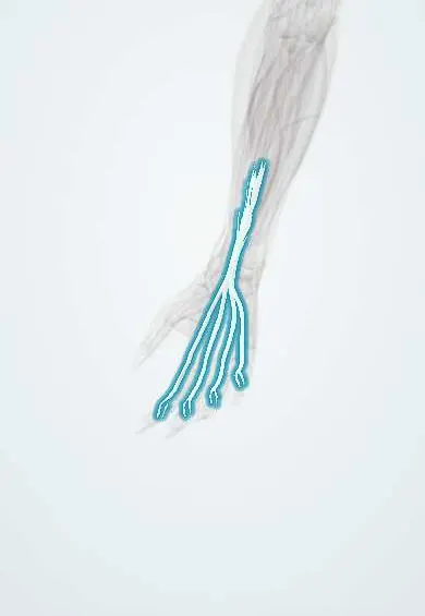
FDS tendons !
The flexor digitorum superficialis tendons run through the wrist into the fingers; they bend the fingers at the middle joints.
Official name: Flexor digitorum superficialis tendons
Opens in the Durgical app, no sign-up needed.
What it is
Four flexor tendons through the carpal tunnel to the middle bones of the fingers; they bend the middle finger joints and can be cut in palm or finger lacerations.
What it does
Bend the fingers at the middle joints.
Key facts
- Attaches
- Four tendons through the carpal tunnel (middle and ring in front of index and little), each splitting at the proximal phalanx to the sides of the middle phalanx
- Function
- Flex the proximal interphalangeal and MCP joints; tested with the other fingers held straight; the FDP tendon passes through its split
- Tight in
- Finger and wrist extension
Relations
- Course
- From the forearm the four tendons pass through the carpal tunnel within the ulnar bursa, fan out in the palm and enter the fibrous sheaths at the A1 pulleys, where they split around the FDP
- Deep
- FDP tendons, behind them in the carpal tunnel and palm until they come through the FDS split over the proximal phalanx
- Superficial
- Median nerve just under the flexor retinaculum in the carpal tunnel; in the palm the superficial palmar arch and the palmar aponeurosis
- Lateral
- FPL tendon, in its own sheath, the most radial tendon in the carpal tunnel
- Medial
- Ulnar nerve and artery, passing over the flexor retinaculum in Guyon's canal
How to find it
With the other fingers held flat on the table, bending one finger at the middle joint shows its FDS working. In the distal forearm the tendons are felt ulnar to the palmaris longus tendon when the fingers flex against resistance.
Examination
- Isolated FDS test: the examiner holds the other fingers fully straight and asks for flexion of the free finger; PIP flexion with a floppy DIP means the FDS is intact, none means it is cut
- Index finger: its FDP often has its own belly, so the standard test is unreliable; a pinch with the PIP bent and the DIP straight shows the FDS instead
- Tenodesis effect: wrist extension normally curls the fingers; a finger that stays straight suggests divided flexor tendons
On imaging
Ultrasound shows tendon continuity during active and passive movement, finds retracted ends before repair and shows tenosynovitis within the sheath.
Clinical notes
- Flexor zones: I distal to the FDS insertion, II from the A1 pulley to the FDS insertion, III palm, IV carpal tunnel, V forearm
- In zone II both tendons lie in the sheath; repair aims to keep them gliding without adhesions, usually with early protected movement
- The ring finger FDS is a common donor in tendon transfers, for example to restore thumb opposition
- 'Spaghetti wrist': deep cuts at the front of the wrist (zone V) often divide several FDS tendons together with the median and ulnar nerves
Anatomical variants
- The little finger FDS is often weak, linked to the ring finger tendon or absent, so a weak isolated test there may be normal
- Accessory slips or bellies can appear in the palm
Memory aid
'Superficialis splits, profundus perforates': the FDS divides so the FDP can pass through it to the fingertip.
Related conditions
Test yourself
Think of the answer, then tap the question to see it.
Where do the FDS tendons insert, and what is Camper's chiasm?
Each splits over the proximal phalanx to let the FDP through; the halves cross again behind it (Camper's chiasm) and insert on the sides of the middle phalanx.
How is an FDS tendon tested in isolation?
The other fingers are held straight, locking their shared FDP; PIP flexion of the free finger with a floppy DIP shows an intact FDS.
What is flexor zone II, and why is it difficult?
From the A1 pulley to the FDS insertion; both flexor tendons share a tight sheath, so repairs tend to form adhesions.
Which FDS tendon is a common donor in tendon transfers?
The ring finger FDS, for example to restore thumb opposition.
Educational information, not medical advice. For any symptom, see a health professional.
Its names
- Official Latin (TA2)
- tendines musculi flexoris superficialis digitorum TA2 2486
- Official Spanish (DeCS)
- Tendones del músculo flexor superficial de los dedos
- English (TA2)
- Flexor digitorum superficialis tendons
Related
- Median nerve
- A1 pulleys
- Middle phalanx, index finger
- Middle phalanx, middle finger
- Middle phalanx, ring finger
- Middle phalanx, little finger
- Proximal phalanx, middle finger
- Proximal phalanx, little finger
Sources: Terminologia Anatomica (TA2); Anatomy, Shoulder and Upper Limb, Hand Flexor Digitorum Superficialis Muscle; Anatomy, Shoulder and Upper Limb, Hand Long Flexor Tendons and Sheaths.
Spotted a mistake?We are always improving for the community. Tell us what is wrong and our team will review it.Report a mistake
Representative model, for educational purposes only.