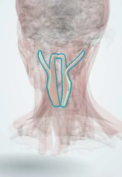
Muscular system · Head and neck
Longitudinal pharynx muscles !
The longitudinal pharynx muscles are long, thin muscles running down inside the throat wall; they lift and shorten the pharynx when swallowing.
Official name: Stylopharyngeus muscle
Opens in the Durgical app, no sign-up needed.
What it is
Long thin muscles running down inside the throat wall.
What it does
Lift and shorten the pharynx when swallowing.
Key facts
- Origin
- Styloid process (stylopharyngeus); palatine aponeurosis (palatopharyngeus)
- Insertion
- Pharyngeal wall and thyroid cartilage
- Action
- Elevate and shorten the pharynx and larynx
- Nerve
- Stylopharyngeus: glossopharyngeal nerve (only muscle of IX); palatopharyngeus: vagus nerve
- Blood supply
- Ascending pharyngeal artery
Relations
- Course
- Stylopharyngeus runs from the inner side of the styloid process down and forward between the internal and external carotid arteries, slips between the superior and middle constrictors and ends on the pharyngeal wall and the thyroid cartilage
- Superficial
- The pharyngeal constrictors, which enclose them
- Deep
- Pharyngobasilar fascia and the mucosa of the pharynx
How to find it
The palatopharyngeal arch is seen through the open mouth behind the tonsil; the salpingopharyngeal fold is seen on nasendoscopy running down from the torus tubarius. The stylopharyngeus is found in dissection between the internal and external carotid arteries.
On imaging
Too thin to separate on routine imaging; on CT or MRI they sit in the pharyngeal wall, inside the constrictors. Their action is judged on videofluoroscopy (modified barium swallow): the pharynx and larynx rise and the pharynx shortens with each swallow. Weak elevation leaves residue in the valleculae and piriform sinuses and can lead to aspiration. Endoscopic study is the bedside option.
Clinical notes
- The glossopharyngeal nerve is traced by following the stylopharyngeus; glossopharyngeal neuralgia gives brief, sharp pain in the throat, tonsil area and ear, set off by swallowing
- Sphincter pharyngoplasty for velopharyngeal insufficiency moves flaps containing the palatopharyngeus into the back wall of the nasopharynx
- A pure lesion of the glossopharyngeal nerve causes almost no visible motor loss, because the stylopharyngeus is its only muscle
Development
The stylopharyngeus comes from the third pharyngeal arch (hence IX); the palatopharyngeus and salpingopharyngeus come from the fourth (hence the vagus nerve).
Test yourself
Think of the answer, then tap the question to see it.
Which three muscles form the longitudinal pharyngeal layer, and what do they do?
Stylopharyngeus, palatopharyngeus and salpingopharyngeus; they lift and shorten the pharynx and larynx during swallowing.
Which nerve supplies the stylopharyngeus, and what is special about it?
The glossopharyngeal nerve; the stylopharyngeus is its only muscle (third pharyngeal arch).
Where does the stylopharyngeus enter the pharynx?
Through the gap between the superior and middle constrictors, with the glossopharyngeal nerve winding round it.
Educational information, not medical advice. For any symptom, see a health professional.
Its names
- Official Latin (TA2)
- musculus stylopharyngeus TA2 2190
- Official Spanish (DeCS)
- Músculos estilofaríngeo y palatofaríngeo
- English (TA2)
- Stylopharyngeus muscle
- Also called
- musculus stylopharyngicus
Related
Sources: Terminologia Anatomica (TA2); Anatomy, Head and Neck, Stylopharyngeus Muscles; Anatomy, Head and Neck, Pharynx.
Spotted a mistake?We are always improving for the community. Tell us what is wrong and our team will review it.Report a mistake
Representative model, for educational purposes only.