Auditory tube !
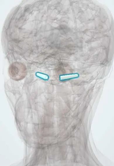
The auditory (Eustachian) tube links the middle ear to the back of the nose and balances pressure behind the eardrum; it is shorter and flatter in children.
Opens in the Durgical app, no sign-up needed.
What it is
A narrow tube from the middle ear to the back of the nose.
What it does
Balances the pressure behind the eardrum; it is short and flat in children, so ear infections are common.
Key facts
- Where
- Middle ear to the nasopharynx (~3.5 cm; shorter, flatter in children)
- Blood supply
- Ascending pharyngeal, middle meningeal arteries
- Nerve
- IX (tympanic plexus) and pharyngeal branch of V2; opened by tensor veli palatini (V3)
- Function
- Equalises middle-ear pressure; drains secretions
Relations
- Course
- From the anterior wall of the middle ear forward, medially and downward to the lateral wall of the nasopharynx, opening behind the inferior nasal concha
- Above
- Canal for the tensor tympani above the bony part
- Behind
- Carotid canal with the internal carotid artery, close to the bony part
- Lateral
- Tensor veli palatini, with the infratemporal fossa beyond
- Below
- Levator veli palatini, under the cartilaginous part; behind the pharyngeal opening, the torus tubarius and the pharyngeal recess (fossa of Rosenmüller)
How to find it
The pharyngeal opening is seen on nasal endoscopy in the lateral wall of the nasopharynx, framed above and behind by the cartilage ridge of the torus tubarius; the adenoids lie on the roof and back wall nearby.
Examination
- Valsalva manoeuvre (blowing against a closed nose and mouth) or Toynbee manoeuvre (swallowing with the nose pinched): visible movement of the drum on otoscopy shows the tube opens
- Tympanometry: type C (negative pressure) suggests tube dysfunction; type B with normal volume suggests fluid behind the drum
On imaging
High-resolution temporal bone CT shows the bony part running forward and medially from the middle ear; the cartilaginous part and its opening beside the torus tubarius show on MRI or nasal endoscopy. A blocked tube shows as fluid in the middle ear and mastoid. In an adult with fluid in one ear only, endoscopy or MRI rules out a nasopharyngeal tumour, often from the fossa of Rosenmüller.
Common problems
- Blocked tube with colds and allergies: fullness and muffled hearing.
- In children it drains poorly, so fluid and infections build up behind the eardrum.
How it is diagnosed
Otoscopy and tympanometry.
Treatment
- Treat the cold or allergy; equalising manoeuvres.
- Ventilation tubes in the eardrum for children with persistent fluid.
Clinical notes
- Otitis media with effusion (glue ear) in children, linked to enlarged adenoids; grommets ventilate the middle ear by bypassing the tube, and adenoidectomy may be added
- Cleft palate: the tensor veli palatini cannot open the tube, so effusion is almost universal
- Barotrauma: on descent in flight or diving a closed tube cannot equalise pressure, causing pain, effusion or haemorrhage behind the drum
- Patulous tube: an abnormally open tube, often after weight loss, causes autophony (hearing one's own voice and breathing loudly)
- The pharyngeal recess behind the torus is the commonest site of nasopharyngeal carcinoma
Development
The tube and the middle ear cavity develop from the first pharyngeal pouch (tubotympanic recess).
Related conditions
- Acute otitis media
- Otitis media with effusion and chronic suppurative otitis media
- Nasopharyngeal carcinoma
Test yourself
Think of the answer, then tap the question to see it.
Which muscle mainly opens the auditory tube, and when?
The tensor veli palatini (V3), helped by the levator veli palatini and salpingopharyngeus, when swallowing or yawning.
Why is otitis media more common in children?
Their auditory tube is shorter, wider and more horizontal: nasopharyngeal secretions reach the middle ear easily and drainage is poor.
What does a one-sided middle ear effusion in an adult raise suspicion of?
A nasopharyngeal tumour blocking the auditory tube; the pharyngeal recess behind the torus tubarius is the commonest site of nasopharyngeal carcinoma.
Which parts of the auditory tube are bony and cartilaginous, and where is it narrowest?
Lateral third bony, medial two thirds cartilaginous; narrowest at the isthmus where the two meet.
What is a patulous auditory tube?
An abnormally open tube, often after weight loss, causing autophony: hearing one's own voice and breathing loudly.
Educational information, not medical advice. For any symptom, see a health professional.
Its names
- Official Latin (TA2)
- tuba auditiva TA2 6926
- Official Spanish (DeCS)
- Trompa Auditiva DeCS D005064
- English (TA2)
- Auditory tube
- Also called
- Eustachian tube · pharyngotympanic tube · tuba pharyngotympanica · tuba Eustachii
Sources: Terminologia Anatomica (TA2); DeCS; Eustachian Tube Dysfunction; Anatomy, Head and Neck, Nasopharynx.
Spotted a mistake?We are always improving for the community. Tell us what is wrong and our team will review it.Report a mistake
Representative model, for educational purposes only.