Muscular system · Head and neck
Pharyngeal constrictors !
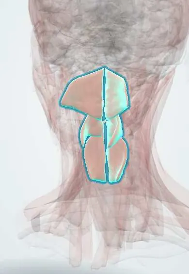
The pharyngeal constrictors are three stacked curved muscles forming the back and sides of the throat; they squeeze in turn to push food down when swallowing.
Official name: Superior pharyngeal constrictor
Opens in the Durgical app, no sign-up needed.
What it is
Three stacked curved muscles that form the back and sides of the throat.
What it does
Squeeze in sequence to push food down when swallowing.
Key facts
- Origin
- Superior: pterygomandibular raphe, medial pterygoid plate; middle: hyoid horns; inferior: thyroid and cricoid cartilages
- Insertion
- Pharyngeal raphe (posterior midline)
- Action
- Squeeze food down in sequence when swallowing
- Nerve
- Vagus nerve via the pharyngeal plexus (inferior also external/recurrent laryngeal)
- Blood supply
- Ascending pharyngeal, inferior thyroid arteries
Relations
- Contents
- Through the gaps: above the superior, the auditory tube and levator veli palatini; between superior and middle, stylopharyngeus and the glossopharyngeal nerve; between middle and inferior, the internal laryngeal nerve and superior laryngeal vessels; below the inferior, the recurrent laryngeal nerve
- Behind
- Buccopharyngeal fascia and the retropharyngeal space, then the prevertebral fascia and the cervical vertebrae
- Deep
- Longitudinal muscles (stylopharyngeus, palatopharyngeus, salpingopharyngeus), the pharyngobasilar fascia above, then the mucosa; the palatine tonsil sits against the superior constrictor
- In front
- Front attachments: pterygomandibular raphe (shared with the buccinator), the horns of the hyoid bone, the thyroid cartilage and the cricoid cartilage
- Lateral
- Parapharyngeal space and the carotid sheath; lower down, the lobes of the thyroid gland
How to find it
Not palpable from outside. A gloved finger in the mouth feels the front of the upper cervical vertebrae through the thin back wall of the pharynx; in dissection the constrictors are seen from behind once the retropharyngeal space is opened.
On imaging
A videofluoroscopic swallow study or a fibre-optic endoscopic evaluation of swallowing (FEES) shows weak pharyngeal squeeze as residue in the valleculae and piriform fossae, and penetration or aspiration into the airway.
Clinical notes
- Curtain sign: with a one-sided vagus nerve or pharyngeal plexus lesion, the back wall of the pharynx moves toward the healthy side on saying 'aah'
- Cricopharyngeal dysfunction: the upper sphincter fails to open fully; a cricopharyngeal bar shows on barium swallow, and it is linked with Zenker diverticulum
- The recurrent laryngeal nerve enters the larynx under the lower edge of the inferior constrictor, behind the cricothyroid joint: a point of risk in thyroid surgery
- Stroke, motor neuron disease and myasthenia gravis commonly weaken the pharyngeal phase, causing dysphagia and aspiration pneumonia
Development
They come from the fourth pharyngeal arch and are supplied by the vagus through the pharyngeal plexus; some texts assign the cricopharyngeus to the sixth arch, matching its supply from the recurrent laryngeal nerve.
Related conditions
Test yourself
Think of the answer, then tap the question to see it.
What is the nerve supply of the pharyngeal constrictors?
The vagus nerve via the pharyngeal plexus; the inferior constrictor also gets the external and recurrent laryngeal nerves.
Which part of the inferior pharyngeal constrictor forms the upper oesophageal sphincter?
The cricopharyngeus, its lowest part; it relaxes as the hyoid and larynx are pulled up and forward.
What is the curtain sign?
With a one-sided vagus nerve or pharyngeal plexus lesion, the back wall of the pharynx moves toward the healthy side on saying «aah».
What passes between the middle and inferior pharyngeal constrictors?
The internal laryngeal nerve and the superior laryngeal vessels.
Where does the recurrent laryngeal nerve enter the larynx relative to the pharyngeal constrictors?
Under the lower edge of the inferior constrictor, behind the cricothyroid joint: a point of risk in thyroid surgery.
Educational information, not medical advice. For any symptom, see a health professional.
Its names
- Official Latin (TA2)
- constrictor superior pharyngis TA2 2179
- Official Spanish (DeCS)
- Músculos constrictores de la faringe DeCS D010609
- English (TA2)
- Superior pharyngeal constrictor
- Also called
- superior constrictor muscle of pharynx · musculus cephalopharyngicus
Related
Sources: Terminologia Anatomica (TA2); DeCS; Anatomy, Head and Neck, Pharynx; Anatomy, Head and Neck, Swallowing; Anatomy of the Human Body (20th ed.): Surface Markings of Special Regions of the Head and Neck (Lea & Febiger (Gray's Anatomy, 1918), 1918) · adapted, Public domain.
Spotted a mistake?We are always improving for the community. Tell us what is wrong and our team will review it.Report a mistake
Representative model, for educational purposes only.