Digestive system · Head and neck
Palatine tonsils !
The palatine tonsils are two oval pads of lymph tissue at the sides of the back of the mouth; they catch germs and swell in tonsillitis.
Official name: Palatine tonsil
Opens in the Durgical app, no sign-up needed.
What it is
Two oval lymph tissue pads at the sides of the back of the mouth.
What it does
Catch germs entering the mouth; they swell in tonsillitis.
Key facts
- Where
- Between the palatoglossal and palatopharyngeal arches
- Blood supply
- Tonsillar branch of the facial artery (main)
- Nerve
- IX (referred ear pain after tonsillectomy)
- Function
- Immune surveillance (Waldeyer's ring)
Relations
- In front
- Palatoglossal arch (anterior pillar)
- Behind
- Palatopharyngeal arch (posterior pillar)
- Lateral
- Capsule and loose peritonsillar tissue, the superior pharyngeal constrictor, then the parapharyngeal space with the glossopharyngeal nerve and the internal carotid artery
- Above
- Soft palate and the supratonsillar fossa, where peritonsillar abscesses often begin
- Below
- Tongue base, with the lingual tonsil
- Medial
- The oropharynx; the tonsillar crypts open on this surface
How to find it
With the tongue held down, each tonsil is seen in its fossa between the two arches; its size is graded by how far it reaches toward the midline (for example on the Brodsky scale, grades 0–4).
On imaging
Contrast CT of the neck separates a peritonsillar abscess (a rim-enhancing collection) from cellulitis and shows spread into the parapharyngeal space; intraoral or neck ultrasound is also used.
Common problems
- Tonsillitis: sore throat, fever and painful swallowing; strep throat needs antibiotics.
- Peritonsillar abscess; large tonsils causing snoring or sleep apnoea in children.
How it is diagnosed
Throat exam and a rapid strep test or culture.
Treatment
- Pain relief; antibiotics for strep.
- Tonsillectomy for frequent infections or sleep apnoea.
Clinical notes
- One-sided enlargement of a tonsil in an adult, especially with ulceration or a neck node, raises suspicion of lymphoma or squamous cell carcinoma (often HPV-related)
- Infectious mononucleosis gives large tonsils with grey exudate and enlarged posterior neck nodes
- Lemierre syndrome: rare spread of a throat infection to the internal jugular vein (septic thrombophlebitis), with septic emboli to the lungs, mostly in young adults
- Lymph drains to the jugulodigastric (tonsillar) node of level II, just below the angle of the mandible; it swells in tonsillitis and is a common first site of spread of tonsil cancer
Development
From the endoderm of the second pharyngeal pouch; the supratonsillar fossa is the remnant of the pouch.
Related conditions
- Tangier disease
- Streptococcal pharyngitis
- Peritonsillar abscess
- Infectious mononucleosis
- Diphtheria
- Head and neck squamous cell carcinoma (oral cavity and oropharynx, HPV)
- Tonsillar hypertrophy and recurrent tonsillitis (post-tonsillectomy care)
- Scarlet fever
- X-linked (Bruton) agammaglobulinaemia
Test yourself
Think of the answer, then tap the question to see it.
What forms the bed of the palatine tonsil, and which nerve lies on it?
The superior pharyngeal constrictor, with the glossopharyngeal nerve on it; the internal carotid artery lies further back and out.
What are the signs of a peritonsillar abscess (quinsy)?
Bulging soft palate, uvula pushed to the other side, trismus and a muffled voice; the pus lies between the tonsil capsule and the constrictor.
Where does post-tonsillectomy bleeding usually come from, and when?
The external palatine (paratonsillar) vein; early in the first 24 hours, or late around 5–10 days when the slough separates.
Why is ear pain common after tonsillectomy?
The palatine tonsils are supplied by the glossopharyngeal nerve (IX), so pain from the tonsil bed is referred to the ear along IX.
What does one-sided tonsil enlargement in an adult raise suspicion of?
Lymphoma or squamous cell carcinoma (often HPV-related), especially with ulceration or a neck node.
Educational information, not medical advice. For any symptom, see a health professional.
Its names
- Official Latin (TA2)
- tonsilla palatina TA2 2853
- Official Spanish (DeCS)
- Tonsila Palatina DeCS D014066
- English (TA2)
- Palatine tonsil
- Also called
- tonsils
Related
Sources: Terminologia Anatomica (TA2); DeCS; Anatomy, Head and Neck, Palatine Tonsil (Faucial Tonsils); Anatomy, Head and Neck: Tonsils; Tratado de anatomía humana (9.ª ed.), tomo II, Libro IV Angiología, sección 5: Linfáticos.
Spotted a mistake?We are always improving for the community. Tell us what is wrong and our team will review it.Report a mistake
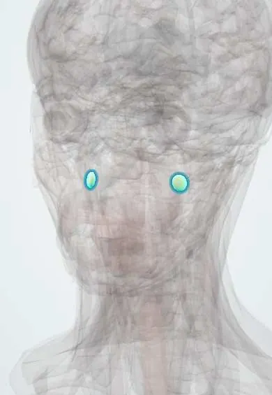
Representative model, for educational purposes only.