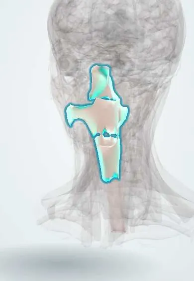
Digestive system · Head and neck
Pharynx !
The pharynx (throat) is the muscular tube behind the nose, mouth and larynx; it carries air to the larynx and food to the oesophagus.
Opens in the Durgical app, no sign-up needed.
What it is
The muscular tube behind the nose, mouth and larynx: naso-, oro- and laryngopharynx.
What it does
Carries air to the larynx and food to the oesophagus.
Key facts
- Where
- Skull base to C6 (cricoid), behind the nose, mouth and larynx
- Blood supply
- Ascending pharyngeal, facial, maxillary, inferior thyroid arteries
- Nerve
- Pharyngeal plexus: motor X; sensory IX (oropharynx), V2 (nasopharynx), X (laryngopharynx)
- Function
- Common passage for air and food
Relations
- In front
- Nasal cavity through the choanae, oral cavity through the oropharyngeal isthmus, larynx through the laryngeal inlet
- Behind
- Retropharyngeal space, prevertebral fascia, longus colli and the bodies of C1–C6
- Lateral
- Parapharyngeal space and carotid sheath (internal carotid artery, internal jugular vein, cranial nerves IX–XII), the styloid muscles; lower down, the thyroid gland lobes
- Above
- Skull base: the sphenoid bone and the basilar part of the occipital bone, through the pharyngobasilar fascia
- Below
- Continues as the oesophagus at the lower border of the cricoid cartilage
How to find it
The oropharynx is seen through the open mouth; the nasopharynx and laryngopharynx are seen with a mirror or a flexible nasendoscope.
On imaging
A lateral neck radiograph shows widened prevertebral soft tissue in retropharyngeal abscess or haematoma; a barium swallow shows a Zenker diverticulum as a pouch behind the junction with the oesophagus; contrast CT maps deep neck abscesses.
Clinical notes
- Zenker diverticulum: dysphagia, regurgitation of undigested food, bad breath and sometimes a gurgling neck swelling; the pouch can be perforated by a blind endoscope or tube
- Nasopharyngeal carcinoma often begins in the lateral recess (fossa of Rosenmüller) behind the auditory tube opening; a one-sided middle ear effusion in an adult calls for a look at the nasopharynx
- Cancers of the laryngopharynx present late, with dysphagia, a neck node or ear pain referred through the vagus nerve
Development
It is the widened head end of the foregut, lined by endoderm; pouches bulge from its side walls and give rise to the tonsils, thymus and parathyroid glands, and branchial cysts and fistulas of the neck come from clefts and pouches that fail to close.
Related conditions
- Streptococcal pharyngitis
- Obstructive sleep apnoea
- Deep neck infection and Ludwig angina
- Caustic ingestion
- Oropharyngeal dysphagia in older adults
- Zenker diverticulum (pharyngo-oesophageal diverticulum)
- Diphtheria
- Common cold (acute viral rhinopharyngitis)
- Head and neck squamous cell carcinoma (oral cavity and oropharynx, HPV)
- Nasopharyngeal carcinoma
- Tonsillar hypertrophy and recurrent tonsillitis (post-tonsillectomy care)
- Juvenile nasopharyngeal angiofibroma
- Acute epiglottitis
Test yourself
Think of the answer, then tap the question to see it.
What are the boundaries of the three parts of the pharynx?
Nasopharynx: skull base to soft palate; oropharynx: soft palate to the epiglottis tip; laryngopharynx: epiglottis to the lower cricoid border (C6).
What is the Killian dehiscence and what forms there?
A weak area at the back of the pharynx between the two parts of the inferior constrictor; a Zenker diverticulum pushes out through it.
Where do fish bones typically lodge in the pharynx, and which nerve lies under that lining?
In the piriform fossae beside the laryngeal inlet; the internal laryngeal nerve runs under their lining.
Where does nasopharyngeal carcinoma often begin, and what adult sign should prompt a look?
In the lateral recess (fossa of Rosenmüller) behind the auditory tube opening; a one-sided middle ear effusion in an adult.
Why is a retropharyngeal abscess dangerous?
The retropharyngeal space runs from the skull base to the upper mediastinum, so an abscess can spread down into the chest.
Educational information, not medical advice. For any symptom, see a health professional.
Its names
- Official Latin (TA2)
- pharynx TA2 2855
- Official Spanish (DeCS)
- Faringe DeCS D010614
- English (TA2)
- Pharynx
- Also called
- throat
Sources: Terminologia Anatomica (TA2); DeCS; Anatomy, Head and Neck, Pharynx; Anatomy of the Human Body (20th ed.): The Digestive Apparatus (Lea & Febiger (Gray's Anatomy, 1918), 1918) · adapted, Public domain; Anatomy of the Human Body (20th ed.): The Branchial Region (Lea & Febiger (Gray's Anatomy, 1918), 1918) · adapted, Public domain.
Spotted a mistake?We are always improving for the community. Tell us what is wrong and our team will review it.Report a mistake
Representative model, for educational purposes only.