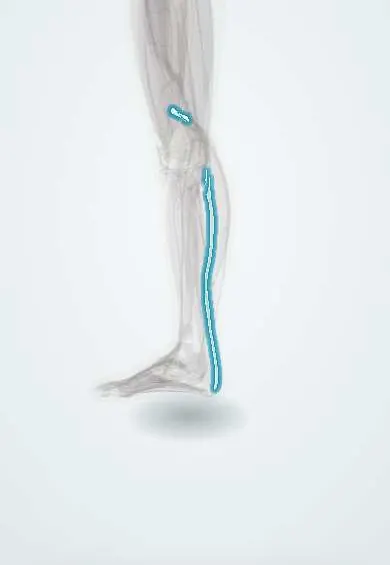
Plantaris tendon !
The plantaris tendon is a long, thin tendon running down the calf to the heel, beside the Achilles tendon.
Official name: Tendon of plantaris
Opens in the Durgical app, no sign-up needed.
What it is
A long, thin tendon running from a small muscle behind the knee down the calf to the heel, beside the Achilles tendon.
What it does
Helps the calf a little; it can be used as a small graft.
Key facts
- Attaches
- Long thin tendon between gastrocnemius and soleus, then along the medial side of the Achilles tendon to the posterior calcaneus (or blending with the Achilles); muscle absent in about 7–20%
- Function
- Weak helper of plantarflexion and knee flexion; often harvested as a free tendon graft; it may stay intact in an Achilles rupture and be mistaken for the Achilles
- Tight in
- Ankle dorsiflexion with the knee extended
Relations
- Course
- From a small belly behind the lateral femoral condyle, the tendon runs obliquely down and inward between the gastrocnemius and the soleus, then along the medial edge of the Achilles tendon to the calcaneus
- Superficial
- Gastrocnemius, mainly its medial head
- Deep
- Soleus
- Lateral
- Achilles tendon, in the lower calf
How to find it
Usually not felt; at surgery it is found as a thin, flat tendon along the medial border of the Achilles tendon and traced upward between the gastrocnemius and the soleus.
Examination
- Thompson (calf squeeze) test, prone: squeezing the calf normally points the foot down; no movement means an Achilles rupture even when the plantaris is intact
On imaging
Ultrasound shows it as a small flat tendon at the medial border of the Achilles tendon; in calf strains it helps tell a plantaris tear from a medial gastrocnemius tear or a venous clot.
Clinical notes
- Harvested through a small incision at the medial heel with a tendon stripper, for ligament reconstructions or tendon augmentation
- Ultrasound can confirm that the tendon is present before it is planned as a graft, because it is missing in some legs
- An isolated plantaris tear leaves a thin band of blood between the gastrocnemius and the soleus on ultrasound and is usually treated without surgery
Anatomical variants
- Its lower attachment varies widely, with at least eight patterns described: on the calcaneus separately, fused with the Achilles tendon, or into nearby fascia
Related conditions
Test yourself
Think of the answer, then tap the question to see it.
In an Achilles rupture with an intact plantaris tendon, what does the Thompson test show?
No foot movement when the calf is squeezed: the Achilles is ruptured even though the thin plantaris cord can be felt.
Why is the plantaris tendon a classic free tendon graft?
It is long, thin and expendable; but the muscle is absent in some people (about 7–20%).
What is the most common cause of 'tennis leg'?
A tear of the medial gastrocnemius; plantaris tears are a minority.
Educational information, not medical advice. For any symptom, see a health professional.
Its names
- Official Latin (TA2)
- musculus plantaris TA2 2663
- Official Spanish (DeCS)
- Tendón del plantar delgado DeCS D013710
- English (TA2)
- Tendon of plantaris
Related
Sources: Terminologia Anatomica (TA2); DeCS; Anatomy, Bony Pelvis and Lower Limb: Calf; Anatomy, Bony Pelvis and Lower Limb: Leg Posterior Compartment.
Spotted a mistake?We are always improving for the community. Tell us what is wrong and our team will review it.Report a mistake
Representative model, for educational purposes only.