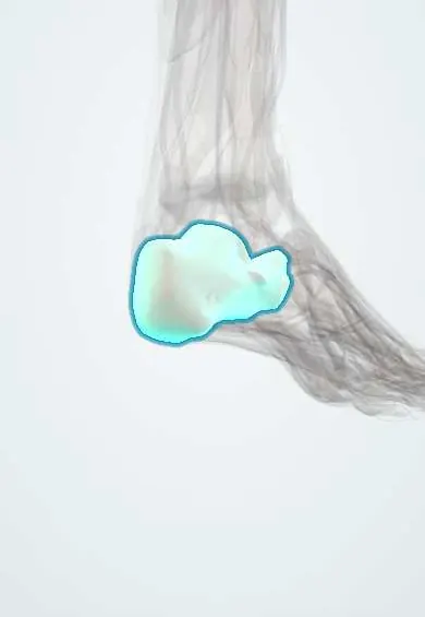
Calcaneus !
The calcaneus, or heel bone, is the largest bone of the foot; it takes weight at each step and anchors the Achilles tendon.
Opens in the Durgical app, no sign-up needed.
What it is
The heel bone, the largest bone in the foot. The talus sits on top of it and the Achilles tendon attaches to its back.
What it does
Takes the impact when the heel strikes the ground and works as a lever for the calf muscles.
Key facts
- Joints with
- Talus (subtalar joint); cuboid (calcaneocuboid joint)
- Landmarks
- Calcaneal tuberosity (Achilles tendon); medial and lateral processes; sustentaculum tali; fibular trochlea; forms sinus tarsi with talus
Relations
- Above
- Talus at the subtalar joint, with the sinus tarsi opening laterally between the two bones
- Behind
- Achilles tendon on the middle of the posterior surface, with the retrocalcaneal bursa between the tendon and the upper bone
- Below
- Plantar fascia, abductor hallucis, flexor digitorum brevis and abductor digiti minimi from the medial and lateral processes; the heel fat pad
- Medial
- Tarsal tunnel: the tibial nerve divides into the medial and lateral plantar nerves beside the posterior tibial vessels; flexor hallucis longus under the sustentaculum
- Lateral
- Fibular tendons, with the fibular trochlea between fibularis brevis (above) and fibularis longus (below); the calcaneofibular ligament; the sural nerve
- In front
- Cuboid at the calcaneocuboid joint; the bifurcate ligament from the anterior process
How to find it
The tuberosity is felt at the back and underside of the heel. About a finger's breadth below the tip of the medial malleolus lies the sustentaculum tali; below the lateral malleolus the fibular trochlea may be felt between the two fibular tendons. The medial process under the heel is the usual point of tenderness in plantar fasciitis.
Examination
- Calcaneal squeeze test: pain on squeezing the heel from both sides suggests a stress fracture, or apophysitis in a child
On imaging
Lateral and axial (Harris) views; Böhler and Gissane angles are measured on the lateral. CT for intra-articular fractures, graded on the posterior facet (Sanders classification). A stress fracture shows as a sclerotic or oedema band across the back of the body, perpendicular to the trabeculae.
Common problems
- Fracture, usually from a fall from a height or a car crash. It can damage the joint under the talus.
- Heel pain, most often from plantar fasciitis at the bottom of the heel.
- Stress fractures in runners and military recruits; a bony bump at the back of the heel (Haglund's deformity) that rubs the Achilles tendon.
How it is diagnosed
A physical exam; X-rays; CT for fractures; MRI for stress fractures.
Treatment
- Many fractures and most heel pain: rest, a boot or cast, then gradual walking and physical therapy.
- Displaced fractures: surgery with plates or screws; sometimes a later fusion of the joint under the talus.
- A bone graft from the patient or a donor is sometimes used to fill gaps in crushed bone.
Clinical notes
- Intra-articular fractures often leave subtalar arthritis and a widened heel that rubs on the fibular tendons
- Anterior process fracture after an inversion injury is easily mistaken for a sprain
- Swelling and fracture blisters often delay surgery for days after the injury
Anatomical variants
- A lucent triangle in the body on the lateral X-ray (pseudocyst) is a normal area with fewer trabeculae
- Calcaneonavicular coalition, seen as the anteater sign on the lateral view
Development
The first tarsal bone to ossify, before birth. The posterior apophysis appears in mid-childhood, normally looks dense and fragmented (not a fracture) and fuses in the mid-teens.
Related conditions
- Plantar Fasciitis
- Achilles tendon rupture
- Diabetic foot
- Osteomyelitis
- Clubfoot (talipes equinovarus)
- Pressure injury (pressure ulcer)
- Marine envenomations and seafood poisoning (ciguatera, scombroid, jellyfish, stingray)
- Mycetoma (actinomycetoma and eumycetoma)
Test yourself
Think of the answer, then tap the question to see it.
What happens to the Böhler angle in a calcaneal fracture that depresses the posterior facet?
It falls below its normal range of about 20–40°; the Gissane angle increases.
After a fall from height with a calcaneal fracture, which other injuries are looked for?
A fracture of the other heel and of the thoracolumbar spine.
Which tendon runs in a groove under the sustentaculum tali?
The flexor hallucis longus tendon.
What is Sever disease?
Calcaneal apophysitis: heel pain in active children at the growth plate where the Achilles tendon pulls.
Educational information, not medical advice. For any symptom, see a health professional.
Its names
- Official Latin (TA2)
- calcaneus TA2 1468
- Official Spanish (DeCS)
- Calcáneo DeCS D002111
- English (TA2)
- Calcaneus
- Also called
- calcaneal bone
Related
- Achilles tendon
- Heel bursae
- Talus
- Cuboid
- Talocalcaneal interosseous ligament
- Spring ligament
- Plantar fascia
- Plantar arteries
Sources: Terminologia Anatomica (TA2); DeCS; Anatomy, Bony Pelvis and Lower Limb: Calcaneus.
Spotted a mistake?We are always improving for the community. Tell us what is wrong and our team will review it.Report a mistake
Representative model, for educational purposes only.