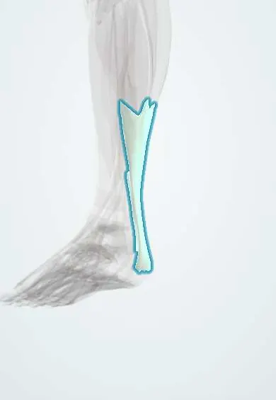
Achilles tendon !
The Achilles tendon is the strongest tendon in the body; it joins the calf muscles to the heel bone and lets the foot push off.
Official name: Calcaneal tendon
Opens in the Durgical app, no sign-up needed.
What it is
The thick tendon at the back of the ankle. It joins the calf muscles (gastrocnemius and soleus) to the heel bone.
What it does
Carries the pull of the calf to the heel, so you can point the foot, push off when walking, run and jump.
Key facts
- Attaches
- Gastrocnemius and soleus (triceps surae) to middle of posterior surface of calcaneus
- Function
- Thickest, strongest tendon; plantarflexion for push-off; ankle jerk reflex (S1–S2)
- Tight in
- Ankle dorsiflexion with knee extended
Relations
- Course
- Forms in mid-calf where the gastrocnemius and soleus tendons merge, narrows to its thinnest a few centimetres above the heel, then widens onto the calcaneus
- Superficial
- Skin and crural fascia; the subcutaneous calcaneal bursa lies between it and the skin at the heel
- Deep
- Kager's fat pad, then the flexor hallucis longus and the deep posterior compartment; at the heel, the retrocalcaneal bursa between it and the posterosuperior calcaneus
- Medial
- Plantaris tendon, which runs along its medial edge and may insert separately on the calcaneus
- Lateral
- Sural nerve and small saphenous vein, which cross from its midline to its lateral side as they descend
How to find it
Lying prone with the feet over the end of the bed, the tendon stands out as a thick cord behind the ankle. In a rupture, a gap is felt a few centimetres above the heel and the resting foot hangs more dorsiflexed than the other.
Examination
- Thompson (Simmonds) test: prone, calf squeezed; no plantarflexion means a complete rupture
- Matles test: prone with both knees bent to 90°; the injured foot falls to neutral or dorsiflexion instead of resting slightly pointed
- Palpable gap along the tendon, best felt early before swelling fills it
- Arc sign: a thickening that moves with the tendon during ankle motion is in the tendon (tendinopathy); one that stays put is in the paratenon
On imaging
Ultrasound is dynamic and measures the gap between the ends with the foot in plantarflexion; MRI shows tendinopathy and partial tears. A lateral X-ray may show loss of Kager's fat triangle in rupture, a Haglund prominence of the calcaneus or insertional calcification.
Common problems
- Tendinopathy: pain, stiffness and thickening from overuse, either in the middle of the tendon or where it attaches to the heel.
- Rupture: a sudden tear, often during a push-off or jump. Many people feel as if they were kicked in the back of the ankle.
- Irritation of the bursa next to the tendon, sometimes with a bony bump on the back of the heel.
How it is diagnosed
A physical exam, including squeezing the calf to see if the foot moves (Thompson test). Ultrasound or MRI shows thickening or a tear.
Treatment
- Tendinopathy: rest from the painful activity, heel lifts and a physical therapy program of gradual strengthening.
- Rupture: a boot or cast with the foot pointed, then physical therapy; or surgery to repair the tendon. The choice depends on the person and the injury.
- Old or large tears: reconstruction, which may use a nearby tendon or a graft from the patient or a donor.
Clinical notes
- Rupture risk rises with fluoroquinolone antibiotics, corticosteroids and pre-existing tendinopathy
- Insertional and midportion tendinopathy are different problems; insertional pain often comes with retrocalcaneal bursitis and a Haglund prominence
- Repair may be open or percutaneous; the sural nerve is at risk with lateral incisions and percutaneous sutures
- Thick, nodular Achilles tendons can be tendon xanthomas of familial hypercholesterolaemia
Anatomical variants
- The plantaris tendon may be absent, join the Achilles tendon or insert separately on the calcaneus
- An accessory soleus muscle can appear as a soft-tissue mass in front of the tendon and cause exertional pain
Related conditions
Test yourself
Think of the answer, then tap the question to see it.
Which muscles form the Achilles tendon, and where does it insert?
The gastrocnemius and soleus (triceps surae); it inserts on the middle of the posterior surface of the calcaneus.
Where do Achilles tendon ruptures and midportion tendinopathy cluster, and why?
About 2–6 cm above the insertion, the area with the poorest blood supply.
How is the Thompson (Simmonds) test done, and what result means a complete Achilles rupture?
With the person prone, the calf is squeezed; no plantarflexion of the foot means a complete rupture.
Why is a complete Achilles tendon rupture often missed?
Active plantarflexion can persist through the deep flexors and fibularis muscles.
Which nerve is at risk with lateral incisions and percutaneous Achilles repair?
The sural nerve, which crosses from the tendon's midline to its lateral side as it descends.
Educational information, not medical advice. For any symptom, see a health professional.
Its names
- Official Latin (TA2)
- tendo calcaneus TA2 2662
- Official Spanish (DeCS)
- Tendón calcáneo DeCS D000125
- English (TA2)
- Calcaneal tendon
- Also called
- Achilles' tendon · tendo Achillis
Related
- Heel bursae
- Calcaneus
- Plantaris tendon
- Soleus tendon
- Sural nerves
- Gastrocnemius (inner head)
- Gastrocnemius (outer head)
- Soleus
Sources: Terminologia Anatomica (TA2); DeCS; Anatomy, Bony Pelvis and Lower Limb: Achilles Tendon.
Spotted a mistake?We are always improving for the community. Tell us what is wrong and our team will review it.Report a mistake
Representative model, for educational purposes only.