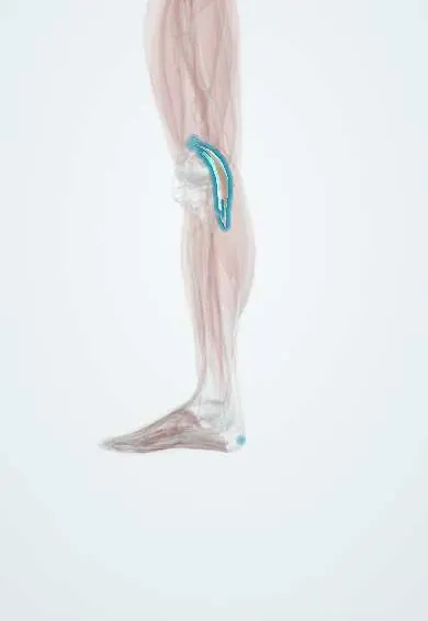
Plantaris !
The plantaris is a small muscle behind the knee with one of the body's longest, thinnest tendons, which runs down to the heel.
Official name: Plantaris muscle
Opens in the Durgical app, no sign-up needed.
What it is
A small muscle behind the knee with a long, thin tendon.
What it does
Helps the calf muscles a little.
Key facts
- Origin
- Lower lateral supracondylar line of femur; oblique popliteal ligament
- Insertion
- Posterior calcaneus, with or medial to calcaneal tendon
- Action
- Weakly assists gastrocnemius (plantar flexes ankle, flexes knee); absent in about 10%
- Nerve
- Tibial nerve (S1–S2)
- Blood supply
- Sural branches of popliteal artery
Relations
- Course
- From above the lateral femoral condyle the belly runs down and medially across the back of the knee; its tendon then passes between gastrocnemius and soleus to reach the medial border of the Achilles tendon
- Superficial
- Lateral head of gastrocnemius at the knee, then the gastrocnemius bellies in the calf
- Deep
- Knee capsule and oblique popliteal ligament, popliteus, then the soleus
- Medial
- Near the heel, the Achilles tendon, beside which it inserts
How to find it
It cannot be felt as a separate muscle. In dissection it is a short belly next to the lateral head of gastrocnemius with a thin, pale, ribbon-like tendon between gastrocnemius and soleus; on ultrasound the tendon is seen beside the medial edge of the Achilles tendon.
On imaging
Ultrasound or MRI shows whether the tendon is present before graft harvest and how it relates to the medial Achilles tendon. In a calf tear, fluid between gastrocnemius and soleus with an intact plantaris tendon points to a gastrocnemius injury.
Clinical notes
- Donor tendon for tendon and ligament reconstructions in the hand and around the ankle
- In some resistant cases of midportion Achilles tendinopathy the plantaris tendon is released or removed
Anatomical variants
- Absence is its best-known variant; how often it is missing depends on the study
- Its insertion varies: on the calcaneus beside the Achilles tendon, merged with that tendon, or into the flexor retinaculum or deep fascia; several insertion types are described
Development
From the ventral (flexor) muscle mass, supplied by the tibial nerve. It is often compared with the palmaris longus of the forearm: both are small, long-tendoned and inconstant.
Memory aid
Nicknamed the "freshman's nerve": its long, pale tendon is easily mistaken for a nerve by new dissectors.
Related conditions
Test yourself
Think of the answer, then tap the question to see it.
Why can the plantaris tendon be used as a free tendon graft?
Its long tendon can be taken with no noticeable loss of function, since the plantaris only weakly assists the gastrocnemius; its presence is checked first, as it may be absent.
Is plantaris rupture the usual cause of «tennis leg»?
No. It was long blamed, but most of these injuries are tears of the medial gastrocnemius head.
Between which muscles does the plantaris tendon run in the calf?
Between the gastrocnemius and the soleus, to the medial side of the Achilles tendon.
Educational information, not medical advice. For any symptom, see a health professional.
Its names
- Official Latin (TA2)
- musculus plantaris TA2 2663
- Official Spanish (DeCS)
- Músculo plantar
- English (TA2)
- Plantaris muscle
Related
Sources: Terminologia Anatomica (TA2); Anatomy, Bony Pelvis and Lower Limb: Calf; Anatomy, Bony Pelvis and Lower Limb: Leg Posterior Compartment.
Spotted a mistake?We are always improving for the community. Tell us what is wrong and our team will review it.Report a mistake
Representative model, for educational purposes only.