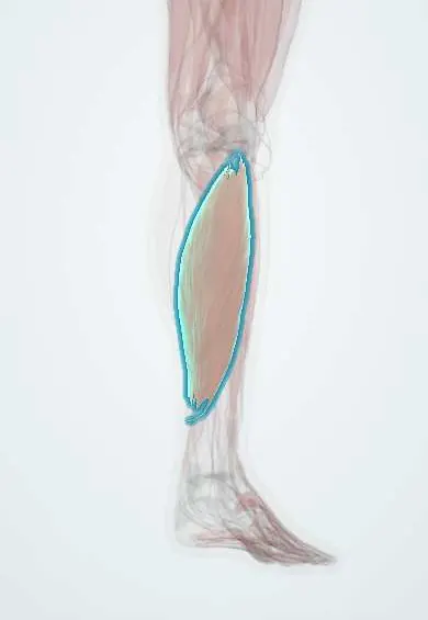
Soleus !
The soleus is a broad, deep calf muscle under the gastrocnemius; it points the foot and keeps the body steady when standing.
Official name: Soleus muscle
Opens in the Durgical app, no sign-up needed.
What it is
A broad, deep calf muscle under the gastrocnemius.
What it does
Points the foot and keeps you steady when standing.
Key facts
- Origin
- Posterior head and upper shaft of fibula; soleal line and medial border of tibia; tendinous arch between them
- Insertion
- Posterior calcaneus via calcaneal (Achilles) tendon
- Action
- Plantar flexes ankle; steadies leg over foot in standing
- Nerve
- Tibial nerve (S1–S2)
- Blood supply
- Posterior tibial, fibular and popliteal (sural) arteries
Relations
- Superficial
- Both heads of gastrocnemius, with the plantaris tendon running between them
- Deep
- Deep transverse fascia and, under it, the deep posterior compartment: tibialis posterior, flexor digitorum longus, flexor hallucis longus, the posterior tibial and fibular vessels and the tibial nerve
- Above
- Popliteus above the soleal line; under its tendinous arch the popliteal artery divides and the tibial nerve continues into the leg
- Below
- Its fibres join the deep surface of the Achilles tendon lower than those of gastrocnemius, often close to the ankle
- Lateral
- Fibularis longus and brevis in the lateral compartment, beyond the posterior intermuscular septum
How to find it
Kneeling on a chair with the knee bent and the heel lifted, the soleus is felt as a firm mass on both sides of the lower calf, below and beside the gastrocnemius bellies and on either side of the Achilles tendon.
Examination
- Silfverskiöld test: dorsiflexion limited with the knee both straight and bent points to soleus or Achilles tightness, not gastrocnemius alone
- Bent-knee (seated) heel raise: weakness or pain with the knee bent singles out the soleus, as in a soleus strain
On imaging
On ultrasound it is the broad muscle deep to the gastrocnemius. Calf compression ultrasound can include its veins: a soleal vein that does not compress and holds echogenic material means a muscular vein thrombosis. MRI shows strains, often along its aponeurosis. An accessory soleus appears on MRI as a muscle mass in front of the Achilles tendon, filling the fat pad there.
Clinical notes
- Superficial posterior compartment syndrome (gastrocnemius, soleus, plantaris): a tense, painful calf with pain on passive ankle dorsiflexion
- The soleus is the classic local muscle flap for defects of the middle third of the tibia (the medial gastrocnemius covers the upper third)
- Soleus strains in runners are deeper and come on more gradually than gastrocnemius tears; bent-knee heel raises reproduce the pain
- The soleal arch is followed in surgery to find the tibial nerve and posterior tibial vessels as they enter the deep posterior compartment
Anatomical variants
- An accessory soleus can appear as a soft-tissue swelling at the back of the ankle, in front of or beside the Achilles tendon, mimicking a tumour and sometimes causing exertional pain
- The arrangement of its fibres varies between people
Development
From the ventral (flexor) muscle mass of the leg; like the gastrocnemius it is supplied by the tibial nerve, mainly through the S1 and S2 roots.
Memory aid
The name comes from Latin solea, a sandal or flat fish: the broad, flat muscle lying under the gastrocnemius.
Related conditions
Test yourself
Think of the answer, then tap the question to see it.
Why does the soleus keep working when the knee is bent?
It is a one-joint muscle that does not cross the knee, so it still acts when the gastrocnemius is slack; bent-knee heel raises single it out.
Why is the soleus linked to deep vein thrombosis?
Its venous sinuses drive the calf muscle pump, and deep vein thrombosis often starts in the soleal veins.
What passes under the tendinous arch of the soleus?
The popliteal vessels and the tibial nerve, entering the deep posterior compartment of the calf.
Which muscle is the classic local flap for defects of the middle third of the tibia?
The soleus; the medial gastrocnemius covers the upper third.
Educational information, not medical advice. For any symptom, see a health professional.
Its names
- Official Latin (TA2)
- musculus soleus TA2 2660
- Official Spanish (DeCS)
- Músculo sóleo
- English (TA2)
- Soleus muscle
Related
Sources: Terminologia Anatomica (TA2); Anatomy, Bony Pelvis and Lower Limb: Calf; Anatomy, Bony Pelvis and Lower Limb: Leg Posterior Compartment.
Spotted a mistake?We are always improving for the community. Tell us what is wrong and our team will review it.Report a mistake
Representative model, for educational purposes only.