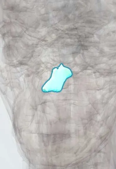
Skeletal system · Head and neck
Vomer !
The vomer is a thin, plough-shaped bone in the lower back part of the nasal septum, the wall that divides the two sides of the nose.
Opens in the Durgical app, no sign-up needed.
What it is
A thin plough-shaped bone in the lower back of the nasal septum.
What it does
Forms part of the wall between the two sides of the nose.
Key facts
- Joints with
- Sphenoid, ethmoid, maxillae, palatines; septal cartilage
- Landmarks
- Lower posterior nasal septum
Relations
- Above
- Rostrum of the sphenoid body between its alae and, in front, the perpendicular plate of the ethmoid bone
- Below
- Nasal crests of the maxillae and palatine bones on the floor of the nose
- In front
- Septal cartilage, set in the angle between its front border and the perpendicular plate
- Behind
- Free border between the choanae, facing the nasopharynx
- Lateral
- Mucosa of each nasal cavity, with the nasopalatine nerve running across it
How to find it
It is not palpable. With a nasal endoscope it is the back part of the septum, and its posterior edge is seen in the midline between the choanae when the nasopharynx is examined.
On imaging
Coronal CT shows the bony septum, deviations and spurs; on axial and sagittal images the vomer is the thin midline plate in front of the nasopharynx.
Clinical notes
- Septoplasty removes or repositions deviated parts of the vomer and septal cartilage while keeping a cartilage strut that supports the nose
- Choanal atresia, a bony or membranous block of the choanae beside the vomer: when bilateral, a newborn has cyanosis that improves on crying
- A nasopalatine nerve block at the incisive papilla numbs the front of the hard palate, the end territory of the nerve that runs along the vomer
Development
Ossifies in membrane from two centres, one on each side of the cartilaginous septum, which fuse; as part of the growing septum, injury in childhood can affect midface growth.
Test yourself
Think of the answer, then tap the question to see it.
What part of the nasal septum does the vomer form, and what does its posterior border divide?
The lower back part of the septum; its free posterior border divides the two choanae in the midline.
Which nerve runs along the vomer, and where is it blocked?
The nasopalatine nerve, in a groove toward the incisive canal; it is blocked at the incisive papilla to numb the front of the hard palate.
How does bilateral choanal atresia beside the vomer present in a newborn?
Cyanosis that improves on crying, because the choanae are blocked.
Educational information, not medical advice. For any symptom, see a health professional.
Its names
- Official Latin (TA2)
- vomer TA2 751
- Official Spanish (DeCS)
- Vómer
- English (TA2)
- Vomer
- Also called
- os vomerale · vomeral bone · vomer bone
Sources: Terminologia Anatomica (TA2); Anatomy, Head and Neck, Nose Bones; Anatomy, Head and Neck, Nasal Cavity.
Spotted a mistake?We are always improving for the community. Tell us what is wrong and our team will review it.Report a mistake
Representative model, for educational purposes only.