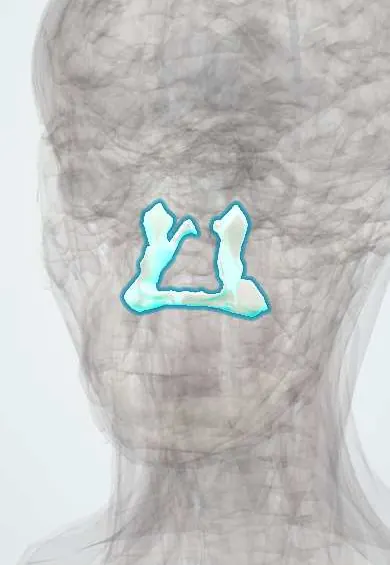
Skeletal system · Head and neck
Palatine bones !
The palatine bones are L-shaped bones at the back of the hard palate; they complete the roof of the mouth and the floor of the nose.
Official name: Palatine bone
Opens in the Durgical app, no sign-up needed.
What it is
L-shaped bones at the back of the hard palate.
What it does
Complete the roof of the mouth and the floor of the nose.
Key facts
- Joints with
- Maxilla, sphenoid, ethmoid, vomer, inferior concha, opposite palatine
- Landmarks
- Horizontal plate (back of the hard palate), greater and lesser palatine foramina; perpendicular plate; pyramidal process; sphenopalatine notch (sphenopalatine foramen); posterior nasal spine
Relations
- In front
- Palatine processes of the maxillae at the transverse palatine suture
- Medial
- Opposite palatine bone at the median palatine suture; the vomer sits on the nasal crest above
- Lateral
- Pterygopalatine fossa, with the greater palatine canal between it and the maxilla
- Behind
- Soft palate, attached to the posterior border of the horizontal plate by the palatine aponeurosis; the posterior nasal spine in the midline; the pterygoid process of the sphenoid bone
- Above
- Floor of the nasal cavity; its small orbital process reaches the back of the orbital floor
How to find it
With the mouth open, the back of the hard palate is felt just in front of the soft palate; the greater palatine foramen lies near the posterior border, medial to the upper third molar, often as a slight soft depression.
On imaging
CT with bone windows shows it best. Coronal images show the horizontal plate at the back of the hard palate and the perpendicular plate in the lateral nasal wall; axial images show the greater palatine foramen beside the upper third molar. In facial trauma, CT shows palatal fractures and the pterygoid plate breaks of Le Fort fractures. MRI looks for tumour spreading along the palatine nerves.
Clinical notes
- Greater palatine nerve block at the foramen numbs the palatal gum and mucosa of the posterior hard palate on that side
- Posterior nosebleeds often come from branches of the sphenopalatine artery entering through the sphenopalatine foramen; endoscopic ligation targets the artery there
- In cleft palate, the gap behind the incisive foramen involves the horizontal plates of the palatine bones and the soft palate
Anatomical variants
- The greater palatine foramen lies most often opposite the upper third molar, but it may sit between the second and third molars or behind the third; texts differ on the proportions
Development
The palatal shelves rise above the tongue and fuse in the midline during the second and third months of development to form the secondary palate; failure of fusion gives cleft palate.
Related conditions
Test yourself
Think of the answer, then tap the question to see it.
Where is the greater palatine foramen of the palatine bone, and what does a block there numb?
At the back corner of the hard palate, medial to the upper third molar; the block numbs the palatal gum and mucosa of the posterior hard palate on that side.
Which artery enters the nose through the sphenopalatine foramen bounded by the palatine bone, and why does it matter?
The sphenopalatine artery; its branches are a common source of posterior nosebleeds, and endoscopic ligation targets it there.
What part of the hard palate do the horizontal plates of the palatine bones form?
The back third of the hard palate; the soft palate attaches to their posterior border.
Educational information, not medical advice. For any symptom, see a health professional.
Its names
- Official Latin (TA2)
- os palatinum TA2 798
- Official Spanish (DeCS)
- Huesos palatinos
- English (TA2)
- Palatine bone
Sources: Terminologia Anatomica (TA2); Anatomy, Head and Neck, Palate; Anatomy, Head and Neck, Skull.
Spotted a mistake?We are always improving for the community. Tell us what is wrong and our team will review it.Report a mistake
Representative model, for educational purposes only.