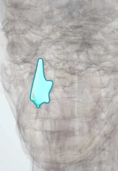
Respiratory system · Head and neck
Nasal septum !
The nasal septum is the wall between the two sides of the nose, cartilage in front and bone behind; when bent (deviated) it can block one side.
Opens in the Durgical app, no sign-up needed.
What it is
The wall between the two sides of the nose, cartilage in front and bone (vomer, ethmoid) behind.
What it does
Divides the airflow; a bent septum can block one side.
Key facts
- Where
- Midline: septal cartilage in front, perpendicular plate of the ethmoid above and behind, vomer below and behind
- Function
- Divides the nasal cavity; Kiesselbach area at its front is the usual nosebleed site; deviation can block one side
Relations
- Above
- Nasal bones and the cribriform plate; the perpendicular plate of the ethmoid bone reaches up to the skull base
- Below
- Nasal crests of the maxillae and palatine bones on the nasal floor; the incisive canal in front
- Lateral
- Each nasal cavity, with the conchae facing it across the airway
- Behind
- Free border of the vomer between the choanae; the sphenoid rostrum above
- In front
- Columella and the medial crura of the major alar cartilages
How to find it
Its lower front edge is seen and felt in the columella between the nostrils; the front of the septum is seen with a speculum and the rest with an endoscope.
Examination
- Inspection after nasal trauma: a boggy swelling of the septum that dents with a probe or cotton bud suggests a septal haematoma
On imaging
Diagnosis is mainly endoscopic; coronal CT shows bony deviation and spurs, usually when sinus surgery is planned.
Common problems
- Deviated septum: blocked breathing on one side, snoring.
- Nosebleeds, most from the front of the septum.
How it is diagnosed
Looking inside the nose; endoscopy.
Treatment
- Nasal sprays.
- Septoplasty to straighten it.
- Pressure on the soft nose for 10-15 minutes, leaning forward, for nosebleeds.
Clinical notes
- Septoplasty corrects a deviated septum while keeping an L-shaped strut of cartilage along the dorsum and the front edge for support
- Septal cartilage is the most used graft material in rhinoplasty
- Loss of septal support after haematoma, abscess or perforation can make the nasal bridge collapse (saddle nose)
Development
Forms from the frontonasal process (merged medial nasal prominences) and grows down to fuse with the palatal shelves; the septal cartilage acts as a growth centre for the midface, so septal surgery in children is conservative.
Related conditions
- Epistaxis (nosebleed)
- Allergic rhinitis
- Leishmaniasis
- Hereditary haemorrhagic telangiectasia (Osler–Weber–Rendu)
- Nasal fracture and septal haematoma
- Granulomatosis with polyangiitis
Test yourself
Think of the answer, then tap the question to see it.
Why can most nosebleeds be seen and treated from the front of the nasal septum?
Kiesselbach (Little) area lies on the front of the cartilaginous septum, within view of a speculum.
Why must a septal haematoma be drained after nasal trauma?
The septal cartilage depends on its perichondrium; without drainage it can die or become infected, leading to a saddle nose.
Which bones and cartilage form the nasal septum?
Septal cartilage in front, the perpendicular plate of the ethmoid above and behind, and the vomer below and behind.
What does septoplasty keep to preserve nasal support?
An L-shaped strut of septal cartilage along the dorsum and the front edge.
What are causes of nasal septum perforation?
Trauma, surgery, repeated cautery, intranasal cocaine use or granulomatosis with polyangiitis.
Educational information, not medical advice. For any symptom, see a health professional.
Its names
- Official Latin (TA2)
- septum nasi TA2 3137
- Official Spanish (DeCS)
- Tabique Nasal DeCS D009300
- English (TA2)
- Nasal septum
Sources: Terminologia Anatomica (TA2); DeCS; Anatomy, Head and Neck, Nasal Cavity; Anatomy, Head and Neck, Nose.
Spotted a mistake?We are always improving for the community. Tell us what is wrong and our team will review it.Report a mistake
Representative model, for educational purposes only.