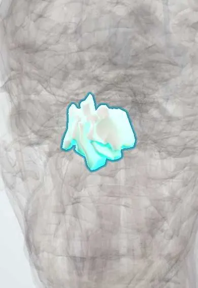
Skeletal system · Head and neck
Ethmoid bone !
The ethmoid is a light, honeycombed bone between the eyes; it forms part of the nose and eye sockets, and its sieve-like roof lets the smell nerves through.
Opens in the Durgical app, no sign-up needed.
What it is
A light, honeycombed bone between the eyes, forming part of the nose and the eye sockets.
What it does
Its sieve-like roof lets the smell nerves through; its air cells lighten the skull.
Key facts
- Joints with
- Frontal, sphenoid, nasals, maxillae, lacrimals, palatines, vomer, inferior conchae
- Landmarks
- Cribriform plate, crista galli, perpendicular plate, ethmoid air cells, superior and middle conchae, orbital plate
Relations
- Above
- Anterior cranial fossa: the olfactory bulbs rest on the cribriform plate on each side of the crista galli, which anchors the falx cerebri; the frontal lobes lie above
- Lateral
- The orbits, separated from the ethmoid cells only by the lamina papyracea; the medial rectus lies against this wall
- Below
- The nasal cavity: the perpendicular plate meets the vomer and septal cartilage in the nasal septum, and the conchae hang from the lateral walls
- In front
- Frontal bone and nasal bones; the frontal sinus drains down through the frontal recess between its anterior cells
- Behind
- Body of the sphenoid bone and the sphenoid sinus
How to find it
It cannot be felt. On coronal CT it sits in the midline between the orbits: cribriform plate and crista galli on top, the air cells on each side and the perpendicular plate below. In endoscopic sinus surgery it is entered through the middle meatus.
On imaging
Coronal CT of the paranasal sinuses (bone window) shows the height and slope of the cribriform plate, the lamina papyracea and the ethmoid cells; it is the standard map before endoscopic sinus surgery.
Clinical notes
- Clear, watery nasal drip after head trauma suggests CSF rhinorrhoea through a cribriform plate fracture, with a risk of meningitis; beta-2 transferrin in the fluid confirms CSF
- Loss of smell after a blow to the head can follow shearing of the olfactory nerve filaments where they pass through the cribriform plate
- In endoscopic sinus surgery the lamina papyracea (orbit) and the skull base above (CSF leak) are the two walls not to breach
- A blind nasal tube is avoided when an anterior skull base fracture is suspected, because it can pass through a broken cribriform plate into the cranial cavity
Anatomical variants
- Concha bullosa: an air cell inside the middle concha; common and usually harmless, though a large one can narrow the middle meatus
- Infraorbital ethmoid (Haller) cells: cells that extend along the orbital floor and can crowd the outflow of the maxillary sinus
- Sphenoethmoidal (Onodi) cell: a posterior ethmoid cell reaching beside or above the sphenoid sinus, next to the optic nerve
- Agger nasi cell: the most anterior ethmoid cell, in front of the attachment of the middle concha; its size shapes the frontal recess
Development
Ossifies in cartilage from three centres: one in each labyrinth before birth and one for the perpendicular plate and crista galli after birth; they fuse in early childhood. The ethmoid cells already exist at birth, so ethmoid sinusitis can occur in infants. (Some texts describe two medial centres, four in all; ossification is complete by about 5–6 years.)
Test yourself
Think of the answer, then tap the question to see it.
What can a fracture of the cribriform plate of the ethmoid bone cause?
Anosmia from torn olfactory filaments and CSF rhinorrhoea from a dural tear, with a risk of meningitis.
Why does ethmoid sinusitis spread to the orbit?
The orbital plate of the ethmoid bone (lamina papyracea) is a paper-thin medial orbital wall separating the ethmoid cells from the orbit.
Which conchae belong to the ethmoid bone, and what drains into the middle meatus?
The superior and middle conchae; the middle meatus receives frontal, maxillary and anterior ethmoid drainage (ostiomeatal complex).
Which test on clear nasal fluid after head trauma confirms CSF from a cribriform plate fracture?
Beta-2 transferrin in the fluid confirms that it is CSF.
Why is a blind nasal tube avoided when an anterior skull base fracture is suspected?
It can pass through a broken cribriform plate of the ethmoid bone into the cranial cavity.
Educational information, not medical advice. For any symptom, see a health professional.
Its names
- Official Latin (TA2)
- os ethmoideum TA2 721
- Official Spanish (DeCS)
- Hueso Etmoides DeCS D005004
- English (TA2)
- Ethmoid bone
- Also called
- ethmoid · ethmoidal bone · os ethmoides
Sources: Terminologia Anatomica (TA2); DeCS; Anatomy, Head and Neck, Ethmoid Bone; Anatomy, Head and Neck, Skull; Anatomía humana (4.ª ed.), tomo 1, cap. 9: Huesos del neurocráneo.
Spotted a mistake?We are always improving for the community. Tell us what is wrong and our team will review it.Report a mistake
Representative model, for educational purposes only.