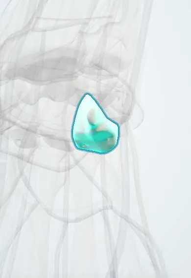
Hamate !
The hamate is a wrist bone on the little-finger side of the second row; its hook can break when a bat, club or racket handle strikes the palm.
Opens in the Durgical app, no sign-up needed.
What it is
A wrist bone on the little-finger side with a hook felt in the palm.
What it does
One of the eight carpal bones that let the wrist bend and carry load to the forearm.
Key facts
- Joints with
- Lunate, triquetrum, capitate, 4th–5th metacarpals
- Landmarks
- Hook (hamulus): lateral wall of Guyon's canal, attachment of the flexor retinaculum
Relations
- In front
- Hook with the flexor retinaculum and the pisohamate ligament; origins of the flexor digiti minimi and opponens digiti minimi
- Medial
- Guyon's canal with the ulnar nerve and ulnar artery, which divide into superficial and deep branches beside the hook
- Lateral
- Capitate; on the radial side of the hook, the carpal tunnel with the FDS and FDP tendons
- Above
- Triquetrum and, in many wrists, the lunate
- Below
- Bases of the 4th and 5th metacarpals, a joint mobile enough to let the ring and little fingers cup the palm
How to find it
The hook is felt by deep pressure in the hypothenar eminence, about 2 cm distal and radial to the pisiform: with the thumb's IP joint over the pisiform and the thumb pointed at the index web, the tip of the thumb lands on it.
Examination
- Hook of hamate pull test: resisted flexion of the ring and little fingers with the wrist in ulnar deviation causes pain when the hook is fractured
On imaging
A carpal tunnel view (wrist extended, beam along the palm) or an oblique view in supination shows the hook; CT is the most reliable. On the PA view the hook shows as a ring (the 'eye'); a missing or blurred ring suggests a fracture.
Clinical notes
- Painful nonunion of the hook is commonly treated by excising the fragment
- Hamate body fractures often accompany fracture-dislocations of the 4th and 5th carpometacarpal joints after a punch
- The deep motor branch of the ulnar nerve can be irritated by a hook fracture or its nonunion, with weakness of the interossei but normal sensation
Anatomical variants
- The hook may ossify from a separate centre that never fuses (os hamuli proprium), which can be mistaken for a fracture; it is often seen on both sides
Development
The second carpal bone to ossify, a few months after the capitate.
Test yourself
Think of the answer, then tap the question to see it.
In which sports is a hook of hamate fracture typical, and how does it present?
Golf, baseball and racket sports, when the handle strikes the palm; it gives deep hypothenar pain and is often missed on routine X-rays.
Which two canals does the hook of the hamate bound?
Guyon's canal on its medial side and the carpal tunnel on its lateral side.
Which tendons can fray or rupture with a nonunion of the hook of the hamate?
The flexor tendons of the ring and little fingers, which glide against the hook.
What is the hook of hamate pull test?
Resisted flexion of the ring and little fingers with the wrist in ulnar deviation; pain suggests a hook fracture.
Educational information, not medical advice. For any symptom, see a health professional.
Its names
- Official Latin (TA2)
- os hamatum TA2 1259
- Official Spanish (DeCS)
- Hueso ganchoso
- English (TA2)
- Hamate
- Also called
- unciform bone
Related
- CMC ligaments
- Dorsal intercarpal ligament
- Dorsal radiocarpal ligament
- Extensor tendons
- FCU tendon
- FDP tendons
- Flexor carpi ulnaris
- Flexor digiti minimi
Sources: Terminologia Anatomica (TA2); Anatomy, Shoulder and Upper Limb, Hand Carpal Bones.
Spotted a mistake?We are always improving for the community. Tell us what is wrong and our team will review it.Report a mistake
Representative model, for educational purposes only.