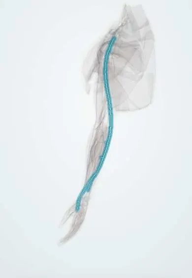
Ulnar nerve !
The ulnar nerve runs behind the inner elbow, the 'funny bone'; it gives feeling to the little and ring fingers and moves most small hand muscles.
Opens in the Durgical app, no sign-up needed.
What it is
The nerve behind the inner elbow bump (the 'funny bone') that runs to the little-finger side of the hand.
What it does
Feeling in the little finger and half the ring finger; powers most small hand muscles.
Key facts
- Roots
- C8–T1 (± C7), medial cord
- Moves
- FCU, FDP (ring, little); hypothenar muscles, interossei, lumbricals 3–4, adductor pollicis, deep FPB, palmaris brevis
- Feeling from
- Little finger and medial half of the ring finger; medial palm and dorsum
Relations
- Course
- From the medial cord it runs medial to the axillary and brachial arteries, pierces the medial intermuscular septum at mid-arm (some texts: passes behind it without piercing it), descends on the medial head of triceps behind the medial epicondyle, enters the forearm between the heads of FCU, runs on FDP under FCU and reaches Guyon's canal lateral to the pisiform
- Lateral
- The ulnar artery, which joins it in the distal two-thirds of the forearm and stays on its lateral side in Guyon's canal
- In front
- Flexor carpi ulnaris in the forearm; at the wrist, the volar carpal ligament and palmaris brevis (roof of Guyon's canal)
- Behind
- The medial epicondyle and the ulnar collateral ligament of the elbow with the joint capsule (floor of the cubital tunnel); flexor digitorum profundus in the forearm
- Medial
- The pisiform at the wrist
How to find it
With the elbow bent, the nerve can be rolled under the fingertip in the groove behind the medial epicondyle; tapping there sends tingling into the little finger. At the wrist it lies deep to the flexor carpi ulnaris tendon, just radial to the pisiform, with the ulnar artery pulse on its radial side.
Examination
- Froment's sign: pinching paper between thumb and index, the thumb IP joint flexes (flexor pollicis longus, median) to make up for a weak adductor pollicis; positive = ulnar motor loss
- Card test and crossed-finger test: failure to hold a card between straight fingers or to cross the middle over the index finger shows weak interossei
- Resisted index abduction (first dorsal interosseous) with a look at the first web space: weakness or wasting is an early motor sign
- Elbow flexion test and Tinel's sign at the cubital tunnel: tingling in the ring and little fingers with the elbow held fully bent supports cubital tunnel syndrome
- Wartenberg's sign: the little finger rests abducted, pulled by extensor digiti minimi against a weak third palmar interosseous
On imaging
Ultrasound shows it in the cubital tunnel behind the medial epicondyle: swelling at or just above the tunnel supports cubital tunnel syndrome, and dynamic scanning in elbow flexion shows it slipping over the epicondyle. At the wrist, Guyon's canal is checked for a ganglion, and CT for a hook of hamate fracture. MRI shows high nerve signal and hand muscle denervation.
Common problems
- Cubital tunnel syndrome: squeezed or stretched behind the elbow; tingling in the ring and little fingers, worse with the elbow bent.
- Compression at the wrist in Guyon's canal (for example cyclists).
- Weakness and wasting of the small hand muscles; clawing of the ring and little fingers.
How it is diagnosed
Tinel at the elbow, the elbow-flexion test, pinch strength (Froment's sign). Nerve conduction studies and EMG.
Treatment
- Avoiding long elbow bending (a night splint) and leaning on the elbow.
- Surgery to release the nerve or move it in front of the elbow (transposition); it is also protected in UCL surgery.
Clinical notes
- Cubital tunnel syndrome is the second commonest entrapment in the upper limb; compression sites include the arcade of Struthers, the medial intermuscular septum, Osborne's ligament and the FCU aponeurosis
- Tardy ulnar palsy: a slowly progressive palsy years after a childhood lateral condyle fracture that left the elbow in cubitus valgus, stretching the nerve
- Medial epicondyle fractures and elbow dislocation can trap or stretch it; it is protected in medial approaches and may be moved forward (anterior transposition)
- Guyon canal zones: 1 before the split (motor and sensory loss), 2 around the deep branch (motor only), 3 around the superficial branch (sensory only)
- Ulnar claw: hyperextended MCP and flexed IP joints of the ring and little fingers; the index and middle are spared because their lumbricals are supplied by the median nerve
Anatomical variants
- Martin-Gruber (forearm) and Riche-Cannieu (palm) anastomoses with the median nerve can keep intrinsic muscles working after an ulnar lesion and confuse nerve studies
- Anconeus epitrochlearis, an accessory muscle bridging the cubital tunnel, can compress the nerve
- In some people the nerve slips over the medial epicondyle on elbow flexion, which can cause friction neuritis
- Its branching pattern inside Guyon's canal varies
Development
Its C8–T1 fibres belong to the postaxial (caudal) border of the limb bud. After the limb turns outward in week 7 that border lies medially, which is why the nerve serves the little-finger side of the forearm and hand.
Memory aid
Interossei, 'PAD and DAB': Palmar ADduct, Dorsal ABduct. Ulnar paradox: the higher the lesion, the straighter the fingers.
Related conditions
Test yourself
Think of the answer, then tap the question to see it.
Where does the ulnar nerve enter the forearm?
Through the cubital tunnel, between the two heads of flexor carpi ulnaris, after passing behind the medial epicondyle.
What is the ulnar paradox?
An ulnar lesion at the elbow claws the ring and little fingers less than one at the wrist, because the FDP to those fingers is lost too.
What does Froment's sign show, and how?
Ulnar motor loss: pinching paper, the thumb IP joint flexes (flexor pollicis longus, median) to make up for a weak adductor pollicis.
What does dorsoulnar hand numbness tell you about the level of an ulnar nerve lesion?
The lesion is above the wrist: the dorsal branch leaves in the forearm, so a Guyon canal lesion spares the dorsum.
What is tardy ulnar palsy?
A slowly progressive ulnar nerve palsy years after a childhood lateral condyle fracture that left cubitus valgus, stretching the nerve.
Educational information, not medical advice. For any symptom, see a health professional.
Its names
- Official Latin (TA2)
- nervus ulnaris TA2 6449
- Official Spanish (DeCS)
- Nervio ulnar
- English (TA2)
- Ulnar nerve
Related
Sources: Terminologia Anatomica (TA2); Anatomy, Shoulder and Upper Limb, Ulnar Nerve; Anatomy, Shoulder and Upper Limb, Hand Guyon Canal; Anatomy of the Human Body (20th ed.): The Branchial Region (Lea & Febiger (Gray's Anatomy, 1918), 1918) · adapted, Public domain; 8.5 Development of the Appendicular Skeleton (OpenStax (Rice University), 2022) · adapted, CC BY 4.0; Anatomía humana (4.ª ed.), tomo 1, cap. 62: Nervios del miembro superior.
Spotted a mistake?We are always improving for the community. Tell us what is wrong and our team will review it.Report a mistake
Representative model, for educational purposes only.