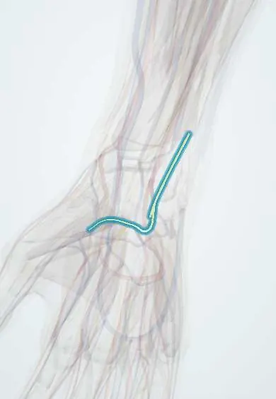
Ulnar nerve in the hand !
In the hand, the ulnar nerve splits in Guyon's canal: a deep branch moves the small hand muscles and a superficial branch gives feeling.
Official name: Deep and superficial branches of the ulnar nerve
Opens in the Durgical app, no sign-up needed.
What it is
The two branches of the ulnar nerve after it passes Guyon's canal at the wrist.
What it does
The deep branch powers the small hand muscles; the superficial branch gives feeling.
Key facts
- Roots
- Ulnar nerve (C8–T1), splits in Guyon's canal
- Moves
- Deep: hypothenar muscles, interossei, lumbricals 3–4, adductor pollicis, deep FPB; superficial: palmaris brevis
- Feeling from
- Superficial branch: palmar little finger and half the ring finger
Relations
- Course
- The deep branch dives between abductor digiti minimi and flexor digiti minimi, pierces opponens digiti minimi, hooks around the hook of the hamate and crosses the palm with the deep palmar arch, deep to the flexor tendons, to end in adductor pollicis and the first dorsal interosseous
- Lateral
- The ulnar artery in Guyon's canal; its deep branch accompanies the deep branch of the nerve
- Medial
- The pisiform at the entry of Guyon's canal
- Deep
- The pisohamate ligament and the flexor retinaculum (floor of Guyon's canal); the hook of the hamate at its exit
- Superficial
- The volar carpal ligament and palmaris brevis (roof of Guyon's canal), under which the superficial branch runs
How to find it
Guyon's canal lies between the pisiform, felt at the base of the hypothenar eminence, and the hook of the hamate: with the thumb's IP crease on the pisiform and the tip aimed at the index finger, the thumb tip presses on the hook. The ulnar artery pulse there lies just radial to the nerve.
Examination
- Froment's sign and card test: positive in a deep branch lesion, because adductor pollicis and the interossei are lost
- Hypothenar bulk and little-finger abduction: normal in a distal deep branch lesion, weak in a lesion at the canal entrance or higher
- Sensation of the palmar little finger (superficial branch) and of the dorsoulnar hand (dorsal branch): both normal in a pure deep branch lesion
On imaging
A carpal tunnel view or CT shows a hook of hamate fracture; ultrasound or MRI shows a ganglion or ulnar artery thrombosis in Guyon's canal.
Clinical notes
- Guyon canal zones: zone 1 before the split (motor and sensory deficit), zone 2 around the deep branch (motor only), zone 3 around the superficial branch (sensory only)
- Causes: ganglion cysts, hook of hamate fractures in golf, baseball and racquet sports, repeated handlebar or tool pressure, and ulnar artery thrombosis (hypothenar hammer syndrome)
- Distal AIN-to-deep-branch nerve transfer can reinnervate the intrinsics after a high ulnar nerve injury
- Clawing is more marked with a lesion here than at the elbow (ulnar paradox), because the ring and little finger FDP still works
Anatomical variants
- The branching inside Guyon's canal varies: the split may be higher or lower, with extra branches or connections
- Riche-Cannieu anastomosis links the deep branch with the recurrent motor branch of the median nerve in the thenar muscles
Memory aid
'PAD and DAB': Palmar interossei ADduct, Dorsal interossei ABduct; both are supplied by the deep branch.
Related conditions
Test yourself
Think of the answer, then tap the question to see it.
Around which bony landmark does the deep branch of the ulnar nerve curve?
The hook of the hamate, after the ulnar nerve splits in Guyon's canal into superficial and deep branches.
What is handlebar palsy of the ulnar nerve in the hand?
Cyclists leaning on handlebars compress the deep branch: a weak, wasted first web space with little or no numbness.
What deficit does each Guyon canal zone give?
Zone 1, before the split: motor and sensory; zone 2, around the deep branch: motor only; zone 3, around the superficial branch: sensory only.
Why are the hypothenar muscles spared by a deep ulnar branch lesion at the hook of the hamate?
Their branches leave earlier; the interossei and adductor pollicis, supplied further on, are weakened.
Educational information, not medical advice. For any symptom, see a health professional.
Its names
- Official Latin (TA2)
- ramus profundus nervi ulnaris TA2 6457
- Official Spanish (DeCS)
- Ramos profundo y superficial del nervio ulnar
- English (TA2)
- Deep and superficial branches of the ulnar nerve
Related
- Pisiform ligaments
- Ulnar artery
- Adductor pollicis
- Dorsal interossei
- Palmar interossei
- Abductor digiti minimi
- Flexor digiti minimi
- Opponens digiti minimi
Sources: Terminologia Anatomica (TA2); Anatomy, Shoulder and Upper Limb, Ulnar Nerve; Anatomy, Shoulder and Upper Limb, Hand Guyon Canal.
Spotted a mistake?We are always improving for the community. Tell us what is wrong and our team will review it.Report a mistake
Representative model, for educational purposes only.