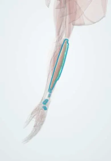
Flexor carpi ulnaris !
The flexor carpi ulnaris runs down the little-finger side of the forearm; it bends the wrist toward the little finger, and the ulnar nerve runs under it.
Opens in the Durgical app, no sign-up needed.
What it is
The forearm muscle on the little-finger side, ending at the pisiform; the ulnar nerve enters the forearm between its two heads.
What it does
Bends the wrist and tilts it toward the little finger.
Key facts
- Origin
- Humeral head: medial epicondyle; ulnar head: olecranon and posterior border of the ulna
- Insertion
- Pisiform, hook of the hamate, base of the 5th metacarpal
- Action
- Flexes and adducts (ulnarly deviates) the wrist
- Nerve
- Ulnar nerve (C7–T1)
- Blood supply
- Ulnar artery (posterior ulnar recurrent)
Relations
- Course
- Runs along the ulnar border of the forearm and becomes tendinous in the distal third; beyond the pisiform it continues as the pisohamate and pisometacarpal ligaments
- Lateral
- Palmaris longus and flexor digitorum superficialis (FDS); in the distal forearm the ulnar artery and ulnar nerve run along its radial side
- Deep
- Flexor digitorum profundus (FDP), with the ulnar nerve lying on it under cover of the muscle along the whole forearm
- Behind
- Its ulnar head attaches by an aponeurosis to the subcutaneous posterior border of the ulna, shared with FDP and the extensor carpi ulnaris (ECU)
How to find it
With the wrist flexed and tilted toward the little finger against resistance, its tendon is felt as a cord just above the pisiform on the inner side of the front of the wrist.
Examination
- Elbow flexion test: holding the elbow fully bent with the wrist extended reproduces tingling in the ring and little fingers in cubital tunnel syndrome; Tinel sign at the tunnel supports it
- Weak wrist flexion toward the little finger with weak FDP to the ring and little fingers places an ulnar lesion at or above the elbow; normal strength with weak hand muscles points to the wrist (Guyon canal)
On imaging
Ultrasound follows the tendon to the pisiform, with the ulnar artery and nerve just radial to it as they enter Guyon's canal. Tendinopathy shows as a thick, hypoechoic tendon. Calcific tendinitis near the pisiform also shows on radiographs as a small calcified density. MRI for tears or unexplained ulnar-sided wrist pain.
Clinical notes
- Cubital tunnel syndrome is the second most common entrapment of the upper limb after carpal tunnel syndrome; surgery releases the arch between the heads, with or without moving the nerve forward
- The interval between flexor carpi ulnaris and FDS is the route to the ulnar artery and nerve in the distal forearm
- Donor for transfer to the finger extensors (extensor digitorum) in radial nerve palsy
Anatomical variants
- An anconeus epitrochlearis, a small muscle bridging the medial epicondyle and olecranon over the cubital tunnel, can compress the ulnar nerve
- Accessory slips of the muscle are rare (case reports)
Related conditions
Test yourself
Think of the answer, then tap the question to see it.
Where does the ulnar nerve enter the forearm in relation to the flexor carpi ulnaris?
Between its two heads, under the aponeurotic arch joining them (Osborne band): the cubital tunnel.
Which bone is a sesamoid in the flexor carpi ulnaris tendon, and what lies beside the tendon at the wrist?
The pisiform; the ulnar artery and nerve lie just lateral to and under the tendon.
What is the elbow flexion test for cubital tunnel syndrome?
Holding the elbow fully bent with the wrist extended reproduces tingling in the ring and little fingers.
How does flexor carpi ulnaris strength help localise an ulnar nerve lesion?
Weak flexor carpi ulnaris and FDP to the ring and little fingers place it at or above the elbow; normal strength with weak hand muscles points to Guyon canal.
Educational information, not medical advice. For any symptom, see a health professional.
Its names
- Official Latin (TA2)
- flexor ulnaris carpi TA2 2483
- Official Spanish (DeCS)
- Músculo flexor ulnar del carpo
- English (TA2)
- Flexor carpi ulnaris
Related
Sources: Terminologia Anatomica (TA2); Anatomy, Shoulder and Upper Limb, Forearm Flexor Carpi Ulnaris Muscle.
Spotted a mistake?We are always improving for the community. Tell us what is wrong and our team will review it.Report a mistake
Representative model, for educational purposes only.