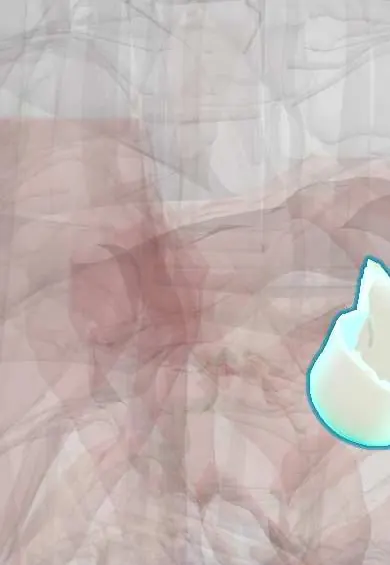
Pulmonary anterior cusp !
The anterior cusp is one of the three flaps of the pulmonary valve, the heart's highest and most forward valve, which opens toward the lungs.
Official name: Anterior semilunar leaflet of pulmonary valve
Opens in the Durgical app, no sign-up needed.
What it is
The front cusp of the pulmonary valve.
What it does
One of the three cusps that seal the pulmonary valve.
Key facts
- Where
- Anterior cusp of pulmonary valve, at top of infundibulum; valve behind sternal end of left 3rd costal cartilage
- Attaches
- Crescent-shaped attachment to pulmonary trunk root; nodule and lunules at free edge
- Function
- Closes in diastole, preventing backflow into right ventricle; valve heard best at left 2nd intercostal space
Relations
- In front
- Front of the chest at the left upper sternal border, partly covered by the edge of the left lung and pleura
- Below
- Smooth infundibulum (conus arteriosus) of the right ventricle
- Above
- Pulmonary trunk
- Behind
- Pulmonary left cusp and pulmonary right cusp, meeting it at the commissures
How to find it
Pulmonic area: left 2nd intercostal space at the sternal border. P2 is heard best here; a loud P2 suggests pulmonary hypertension.
On imaging
Echocardiography: parasternal short axis at the base shows the valve and trunk, with the anterior cusp nearest the chest wall; Doppler measures the stenotic gradient. Cardiac MRI measures regurgitation after repair.
Common problems
- Pulmonary stenosis, usually from birth; often treated with a balloon.
- Leak after childhood repair of congenital heart disease.
How it is diagnosed
Echocardiogram; cardiac MRI.
Treatment
- Balloon valvuloplasty or valve replacement (surgical or by catheter).
Clinical notes
- Pulmonary stenosis is part of tetralogy of Fallot and is common in Noonan syndrome (thick, dysplastic valve)
- Ross procedure: the person's own pulmonary valve replaces the diseased aortic valve and a donor valve takes its place
- Transcatheter pulmonary valve implantation is used for failing valves or conduits after congenital repair
Development
It forms from the anterior cushion of the outflow tract, which stays whole; the other two pulmonary cusps come from halves of the lateral cushions shared with the aorta.
Related conditions
Test yourself
Think of the answer, then tap the question to see it.
Where is the pulmonary valve heard, and which component of the second heart sound does it give?
At the left 2nd intercostal space by the sternum; its closure gives P2, the second and softer part of S2. A loud P2 suggests pulmonary hypertension.
How does normal splitting of S2 change with inspiration, and what does a fixed split suggest?
It widens on inspiration because P2 comes later; a fixed split suggests an atrial septal defect.
What is the Ross procedure?
The person's own pulmonary valve replaces the diseased aortic valve, and a donor valve takes the pulmonary position.
Educational information, not medical advice. For any symptom, see a health professional.
Its names
- Official Latin (TA2)
- valvula semilunaris anterior valvae trunci pulmonalis TA2 4012
- Official Spanish (DeCS)
- Valva semilunar anterior de la válvula pulmonar DeCS D011664
- English (TA2)
- Anterior semilunar leaflet of pulmonary valve
- Also called
- non-adjacent leaflet of pulmonary valve · anterior semilunar cusp of pulmonary valve · velum semilunare sinistrum
Related
Sources: Terminologia Anatomica (TA2); DeCS; Anatomy, Thorax, Heart Pulmonic Valve; Anatomy of the Human Body (20th ed.): Development of the Vascular System (Lea & Febiger (Gray's Anatomy, 1918), 1918) · adapted, Public domain.
Spotted a mistake?We are always improving for the community. Tell us what is wrong and our team will review it.Report a mistake
Representative model, for educational purposes only.