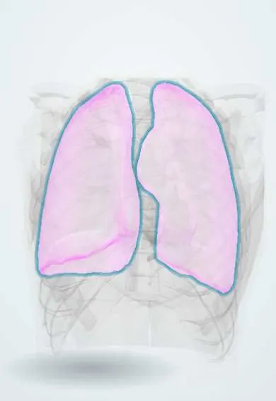
Pleura !
The pleura is the thin double lining around each lung and inside the chest wall; it lets the lungs glide and expand smoothly as we breathe.
Opens in the Durgical app, no sign-up needed.
What it is
A thin, two-layered lining: one layer covers each lung, the other lines the inside of the chest wall, with a thin film of fluid between them.
What it does
Lets the lungs glide as you breathe and keeps them stuck to the chest wall so they expand with it.
Key facts
- Where
- Visceral pleura on lungs; parietal pleura lines cavity: costal, mediastinal, diaphragmatic and cervical parts
- Attaches
- Layers meet at lung hilum and pulmonary ligament; cervical pleura rises above clavicle under suprapleural membrane
- Function
- Fluid film lets lungs glide and couples them to chest wall; parietal pleura pain-sensitive (intercostal and phrenic nerves)
Relations
- Superficial
- The parietal pleura lies on the endothoracic fascia, deep to the innermost intercostal muscles, ribs and intercostal vessels and nerves
- Above
- Cervical pleura under the suprapleural membrane, with the subclavian artery and vein, the brachial plexus and the stellate ganglion
- Medial
- Mediastinal pleura on the pericardium, phrenic and vagus nerves, great vessels and esophagus; it sleeves the root of the lung and hangs below it as the pulmonary ligament
- Below
- Diaphragmatic pleura on the diaphragm; laterally it folds onto the chest wall at the costodiaphragmatic recess
- Contents
- The pleural cavity: a potential space with a thin film of serous fluid between the visceral and parietal layers
How to find it
Lower limits: 8th rib in the midclavicular line, 10th in the midaxillary line, 12th beside the spine. In front, the two pleurae meet behind the sternum from about the 2nd to the 4th costal cartilage, then the left one moves away at the cardiac notch. Medially it dips below the 12th rib, so it can be opened in posterior kidney approaches.
Examination
- Percussion: stony dullness over a pleural effusion; increased resonance over a pneumothorax
- Tactile fremitus and breath sounds: reduced or absent over fluid or air in the pleural cavity
- Pleural friction rub: a creaking sound with each breath, from inflamed pleural layers rubbing together
On imaging
Upright frontal X-ray: a blunted costophrenic angle with a curved meniscus means fluid; a lateral view shows smaller amounts in the posterior recess. Pneumothorax: a thin visceral pleural line with no lung markings beyond it; when lying flat, air collects in front (deep sulcus sign). Ultrasound shows fluid well and loss of lung sliding in pneumothorax.
Common problems
- Pleural effusion: fluid collects between the layers (from heart failure, infection, cancer).
- Pneumothorax: air gets in and the lung collapses.
- Pleurisy: inflammation causing sharp pain with breathing.
How it is diagnosed
Chest X-ray, ultrasound, CT; testing the fluid.
Treatment
- Draining fluid or air with a needle or a chest tube; treating the cause.
Clinical notes
- Tension pneumothorax: air trapped under pressure pushes the mediastinum and trachea to the other side and cuts venous return; it is a clinical diagnosis needing immediate decompression
- Needle decompression is classically in the 2nd intercostal space, midclavicular line; current trauma teaching in adults often prefers the 4th or 5th space just in front of the midaxillary line
- Thoracentesis enters just above the upper border of a rib, because the intercostal vein, artery and nerve run in the groove under the rib above
- Chest drains go in the safe triangle: lateral edge of the pectoralis major, front edge of the latissimus dorsi and a line at the level of the nipple (about the 5th intercostal space), apex below the axilla
Development
The pleural cavities come from the intraembryonic coelom, closed off from the pericardial cavity by the pleuropericardial folds and from the peritoneal cavity by the pleuroperitoneal membranes. Failure of closure, usually on the left, causes a posterolateral (Bochdalek) diaphragmatic hernia.
Memory aid
6, 8, 10 for the lung and 8, 10, 12 for the pleura: midclavicular, midaxillary and paravertebral lines.
Related conditions
- Dengue
- Systemic lupus erythematosus
- Parapneumonic effusion and empyema
- Pneumoconioses (silicosis, asbestosis, coal workers' pneumoconiosis)
- Malignant pleural mesothelioma
- Familial Mediterranean fever and other hereditary autoinflammatory syndromes
- Major trauma (polytrauma): ATLS primary survey
- Penetrating trauma (stab and gunshot wounds)
- Pulmonary contusion
- Pneumothorax
- Pleural effusion
- Haemothorax
Test yourself
Think of the answer, then tap the question to see it.
What are the lower limits of the lung and of the pleura?
Lung at the 6th, 8th and 10th ribs, pleura at the 8th, 10th and 12th (midclavicular, midaxillary, beside the spine): about two ribs lower.
How high does the cervical pleura rise, and what puts it at risk?
About 2 to 3 cm above the medial third of the clavicle; a subclavian line or a deep wound at the root of the neck can puncture it.
Where is pain from the central diaphragmatic pleura felt, and why?
At the shoulder tip, because it is supplied by the phrenic nerve (C3 to C5); the costal pleura gives chest wall pain.
Why is thoracentesis done just above the upper border of a rib?
The intercostal vein, artery and nerve run in the groove under the rib above.
What are the boundaries of the safe triangle for chest drains?
Lateral edge of the pectoralis major, front edge of the latissimus dorsi and a line at nipple level (about the 5th intercostal space), apex below the axilla.
Educational information, not medical advice. For any symptom, see a health professional.
Its names
- Official Latin (TA2)
- pleura; pleura visceralis; pleura parietalis TA2 3322
- Official Spanish (DeCS)
- Pleura DeCS D010994
- English (TA2)
- Pleura
- Also called
- pulmonary pleura
Related
Sources: Terminologia Anatomica (TA2); DeCS; Anatomy, Thorax, Pleurae; Anatomy, Thorax, Lung Pleura And Mediastinum.
Spotted a mistake?We are always improving for the community. Tell us what is wrong and our team will review it.Report a mistake
Representative model, for educational purposes only.