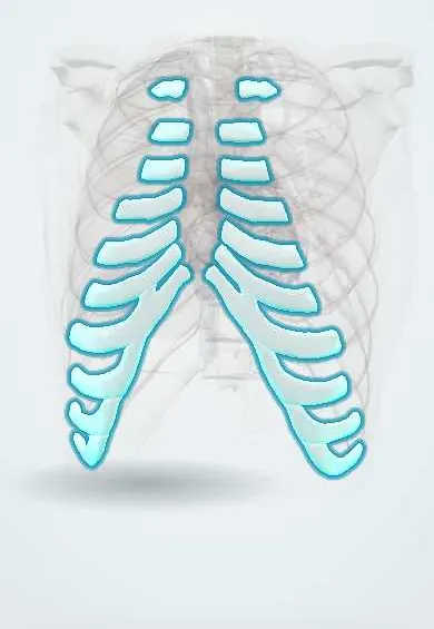
Costal cartilages !
The costal cartilages join the ribs to the breastbone at the front; their flexibility lets the chest expand with each breath.
Official name: Costal cartilage
Opens in the Durgical app, no sign-up needed.
What it is
Bars of cartilage that join the front ends of the upper ten ribs to the sternum or to the cartilage above.
What it does
Give the rib cage its springiness, so the chest can expand when you breathe in.
Key facts
- Where
- Hyaline cartilage at anterior ends of ribs 1–10; ribs 11–12 have only cartilage tips
- Attaches
- Ribs 1–7 to sternum (true ribs); 8–10 to cartilage above, forming costal margin
- Function
- Elasticity of rib cage for breathing; 1st sternocostal joint is a synchondrosis
Relations
- In front
- Pectoralis major over the upper cartilages; rectus abdominis attaches to cartilages 5–7
- Behind
- Internal thoracic artery and veins with the parasternal lymph nodes, on the transversus thoracis; then endothoracic fascia and pleura
- Below
- The lower cartilages give origin to the costal part of the diaphragm and to transversus abdominis on their inner surface
- Medial
- Sternum: cartilages 1–7 reach it directly; 8–10 join the cartilage above to form the costal margin
How to find it
The costal margin is felt along the lower edge of the chest from the xiphoid to the lateral side. The 2nd cartilage is found beside the sternal angle and the others are counted down from it, finger in each space; in thin people the costochondral junctions are felt as a slight step.
Examination
- Palpation of each sternocostal and costochondral junction: reproduction of the person's pain supports costochondritis, but cardiac and other causes of chest pain are excluded first
- Hooking manoeuvre: the examiner curls the fingers under the lower costal margin and pulls forward; pain or a click suggests slipping rib syndrome of cartilages 8–10
On imaging
Cartilage is not seen on plain radiographs unless it calcifies, which is common with age and can mimic lung nodules. CT shows costochondral fractures, Tietze swelling and, in pectus excavatum, the depth of the deformity (Haller index).
Common problems
- Costochondritis: tender, sharp chest-wall pain where the cartilage meets the sternum; it is harmless but can feel like heart pain.
How it is diagnosed
Exam and X-ray; CT finds small fractures and lung injury.
Treatment
- Pain control and deep-breathing exercises to prevent pneumonia; surgical plating for flail chest.
Clinical notes
- Flail chest: when segments of adjacent ribs or cartilages are broken in two places, the loose segment moves inward on inspiration (paradoxical movement); anterior flail can follow costochondral separation or CPR
- Rib cartilage is a common graft source for nasal, ear and airway reconstruction
- In rickets the costochondral junctions enlarge into a row of bumps (rachitic rosary)
- Pectus excavatum can compress the heart and is linked with connective tissue disorders such as Marfan syndrome; it may be corrected surgically, and carinatum is often treated with external bracing
Development
They are the front ends of the embryonic rib models, grown from the costal processes of the thoracic vertebrae, that never turn into bone. They stay hyaline cartilage, which keeps the chest wall springy; the 1st cartilage is often the first to calcify in adults.
Related conditions
Test yourself
Think of the answer, then tap the question to see it.
What type of joint is the 1st sternocostal joint, and what type are joints 2–7?
The 1st is a synchondrosis; joints 2–7 are synovial. The costochondral junctions have no joint cavity.
How does Tietze syndrome differ from costochondritis?
Tietze syndrome is a painful, visible swelling, usually of a single 2nd or 3rd cartilage; costochondritis is tenderness of several upper cartilages without swelling.
Which costal cartilages form the costal margin?
Cartilages 8–10, each joining the cartilage above; cartilages 1–7 reach the sternum directly.
What does the hooking manoeuvre test for?
Slipping rib syndrome of cartilages 8–10: fingers curled under the lower costal margin pull forward, and pain or a click is a positive result.
What causes pectus excavatum and pectus carinatum?
Overgrowth of the costal cartilages, which pushes the sternum in (excavatum) or out (carinatum).
Educational information, not medical advice. For any symptom, see a health professional.
Its names
- Official Latin (TA2)
- cartilago costalis TA2 1140
- Official Spanish (DeCS)
- Cartílago costal DeCS D066186
- English (TA2)
- Costal cartilage
Sources: Terminologia Anatomica (TA2); DeCS; Anatomy, Thorax, Ribs; Anatomy, Thorax, Sternum.
Spotted a mistake?We are always improving for the community. Tell us what is wrong and our team will review it.Report a mistake
Representative model, for educational purposes only.