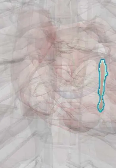
Tetralogy of Fallot !
Tetralogy of Fallot is a congenital heart defect with four features, including a hole between the ventricles, that makes a child's skin bluish.
Updated 2026-10-29
Structures involved
Tap one to see what it is and open it in 3D.
- Interventricular septum
- Right ventricle
- Pulmonary trunk
- Ascending aorta
- Pulmonary anterior cusp
- Pulmonary arteries
- Left ventricle
- Aortic arch
- Subclavian artery
- Thymus
In embryology
- Neural crest cells and their derivatives
- 22q11.2 deletion (DiGeorge): the third and fourth pouches
- Outflow tract: the spiral aorticopulmonary septum and neural crest
- Tetralogy of Fallot
- Isotretinoin and other retinoids
In the clinical exam
In physiology
Why it happens
What it is
A cyanotic congenital heart defect with four features: a ventricular septal defect, right ventricular outflow obstruction, an aorta that overrides the septum and right ventricular hypertrophy. It is the commonest cyanotic heart defect after the newborn period.
Who gets it
The most frequent cyanotic congenital heart disease. It is associated with 22q11.2 deletion (DiGeorge syndrome) and trisomy 21. Thanks to surgery in infancy, a growing number of adults live with repaired tetralogy.
Causes
- Anterior and upward deviation of the outlet (infundibular) septum during development, which produces the four features
- 22q11.2 deletion, trisomy 21 and other genetic syndromes
- In most cases no single cause is found
Risk factors
- Maternal diabetes or uncontrolled phenylketonuria
- Maternal rubella or alcohol use in pregnancy
- Exposure to retinoids in pregnancy
- Family history of congenital heart disease
Pathophysiology
Outflow obstruction raises right ventricular pressure, so deoxygenated blood crosses the large defect into the overriding aorta (right-to-left shunt) and causes cyanosis in proportion to the obstruction. In a tet spell, the obstruction worsens or systemic resistance falls, the shunt increases and hypoxia deepens. Squatting raises systemic resistance and reduces the shunt.
Classification
- «Pink» tetralogy: mild obstruction and little cyanosis
- Classic tetralogy with pulmonary stenosis
- Tetralogy with pulmonary atresia: duct-dependent
- Tetralogy with absent pulmonary valve: rare
Symptoms and signs
- Cyanosis, often increasing over the first months
- Harsh ejection murmur at the upper left sternal border from the outflow obstruction, with a single S2
- Tet spells during crying or feeding: irritability, deep fast breathing and deeper cyanosis
- Squatting after exertion in older children
- Clubbing and polycythaemia if not repaired
Diagnosis
- Clinical
- Cyanosis with an ejection murmur at the upper left sternal border that softens during a spell, and a single S2.
- Tests
- Low pulse oximetry; ECG with right axis deviation and right ventricular hypertrophy; blood count may show polycythaemia; genetic testing for 22q11.2 deletion.
- Imaging
- Echocardiography confirms the diagnosis. Chest X-ray shows a boot-shaped heart with reduced lung markings. CT, MRI or catheterisation define the pulmonary arteries and coronaries; MRI follows the right ventricle after repair.
Differential diagnosis
- Transposition of the great arteries
- Pulmonary atresia with intact septum
- Tricuspid atresia
- Truncus arteriosus
- Persistent pulmonary hypertension of the newborn
- Eisenmenger syndrome
Goal of treatment
Relieve cyanosis and spells, perform complete repair in infancy, then follow for life to manage pulmonary regurgitation and arrhythmias.
Treatment
- Acute
- Tet spell: calm the child, knee-to-chest position and oxygen, then intravenous fluids, opioid sedation, beta-blockers and alpha-agonist vasopressors to raise systemic resistance.
- Acute
- In newborns with severe obstruction or pulmonary atresia, prostaglandin E1 keeps the ductus open.
- Surgical
- Complete repair, usually in the first year: patch closure of the ventricular septal defect and relief of the outflow obstruction.
- Procedure
- When complete repair must wait: a systemic-to-pulmonary shunt (modified Blalock-Taussig) or catheter stenting of the duct or outflow tract.
- Follow-up
- Lifelong specialist follow-up, with pulmonary valve replacement (surgical or transcatheter) for severe regurgitation and rhythm monitoring.
Complications
- Hypoxic (tet) spells
- Polycythaemia, stroke and brain abscess in unrepaired disease
- Infective endocarditis
- After repair: pulmonary regurgitation and right ventricular dilatation
- Ventricular arrhythmias and sudden death
- Residual defect or obstruction, heart block
Prognosis
Without treatment, survival into adulthood is uncommon. After repair in infancy most patients reach adulthood with a good quality of life, but many need reinterventions and lifelong follow-up.
Test yourself
Think of the answer, then tap the question to see it.
What are the four features of tetralogy of Fallot?
Ventricular septal defect, right ventricular outflow obstruction, overriding aorta and right ventricular hypertrophy.
What is the classic chest X-ray sign of tetralogy of Fallot?
A boot-shaped heart (coeur en sabot) with reduced lung markings.
Why does squatting help a child with tetralogy of Fallot?
It raises systemic vascular resistance, which reduces the right-to-left shunt.
Why does the murmur get softer during a tet spell?
The murmur comes from the outflow obstruction; during a spell less blood crosses it.
Which genetic deletion is associated with tetralogy of Fallot?
The 22q11.2 deletion (DiGeorge syndrome).
Related conditions
- Ventricular septal defect
- Patent ductus arteriosus
- Atrial septal defect
- Heart failure
- Ischaemic stroke
- Infective endocarditis
Educational information, not medical advice. For any symptom, see a health professional.
Sources: Tetralogy of Fallot (StatPearls (NCBI Bookshelf), 2024).
Spotted a mistake?We are always improving for the community. Tell us what is wrong and our team will review it.Report a mistake
Representative model, for educational purposes only.