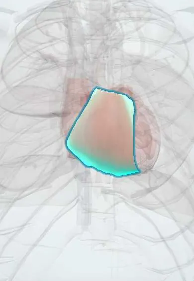
Right ventricle !
The right ventricle is the lower right chamber of the heart, just behind the breastbone; it pumps blood to the lungs to pick up oxygen.
Opens in the Durgical app, no sign-up needed.
What it is
The lower right chamber, a thin-walled pump wrapped around the front of the left ventricle.
What it does
Pumps blood through the pulmonary valve into the lungs, where it picks up oxygen. It works at low pressure.
Key facts
- Where
- Forms most of anterior (sternocostal) surface; trabeculated inflow, smooth outflow (conus arteriosus); moderator band
- Blood supply
- Right coronary artery (right marginal branch); anterior branches of LAD
- Nerve
- Cardiac plexus: sympathetic (T1–T4) speeds and strengthens; vagus nerves slow
- Function
- Pumps deoxygenated blood through pulmonary valve into pulmonary trunk at low pressure; thin wall
Relations
- In front
- Body of the sternum and the left costal cartilages, partly covered by the thin edge of the left lung and pleura
- Behind
- Interventricular septum, with the left ventricle behind and to the left
- Above
- Pulmonary trunk rising from the infundibulum; the right coronary artery runs in the atrioventricular groove along its right upper edge
- Below
- Diaphragm (central tendon), forming a narrow strip of the diaphragmatic surface; below the diaphragm lie the left lobe of the liver and the stomach, passed near by a subxiphoid needle or probe aimed at the heart
- Contents
- Coarse trabeculae carneae, the septomarginal trabecula (moderator band) carrying the right bundle branch, and the right ventricle papillary muscles
How to find it
It sits behind the lower sternum and the left parasternal region. A sustained lift felt with the heel of the hand at the left lower sternal border (parasternal heave) suggests an enlarged or thickened right ventricle. The tricuspid area is the left lower sternal border, 4th to 5th intercostal space.
On imaging
Lateral chest X-ray: an enlarged right ventricle fills the space behind the sternum. Echocardiography: the apical four-chamber view compares its size with the left ventricle; in the parasternal short axis, pressure or volume overload flattens the septum into a D-shaped left ventricle.
Common problems
- Strains and fails when lung pressure is high (pulmonary hypertension, a large pulmonary embolism).
- Can be damaged by an inferior heart attack.
How it is diagnosed
Echocardiogram; right-heart catheterization measures lung pressures.
Treatment
- Treat the cause (lung disease, clots); medicines that lower lung pressure.
Clinical notes
- Pulmonary hypertension and chronic lung disease thicken and dilate it (cor pulmonale); a large pulmonary embolism dilates it suddenly
- Suspected right ventricular infarct with an inferior infarct: right-sided chest leads (V4R) help show it, and these patients depend on adequate filling pressure
- In tamponade its free wall collapses in early diastole on echo, because it is the lower-pressure ventricle
Development
Its trabeculated body comes mostly from the bulbus cordis and its smooth outflow from the conus; forward displacement of the outflow septum underlies tetralogy of Fallot.
Related conditions
- Beriberi (thiamine deficiency)
- Ventricular tachycardia
- Dilated cardiomyopathy
- Long QT syndrome and torsades de pointes
- Constrictive pericarditis
- Transposition of the great arteries
- Pulmonary hypertension and cor pulmonale
- Arrhythmogenic right ventricular cardiomyopathy
- Brugada syndrome
- Eisenmenger syndrome
- Ebstein anomaly
- Blunt cardiac injury
- Myocardial Infarction (Heart Attack)
- Heart failure
- Pulmonary embolism
- Cardiac tamponade
- Ventricular septal defect
- Tetralogy of Fallot
Test yourself
Think of the answer, then tap the question to see it.
Why is the right ventricle the chamber most often hit in penetrating chest wounds?
It lies directly behind the sternum and forms most of the anterior (sternocostal) surface of the heart.
Which structure in the right ventricle carries the right bundle branch?
The septomarginal trabecula (moderator band), which runs from the septum to the anterior papillary muscle.
What is the classic picture of right ventricular infarction, and which artery is usually occluded?
Low blood pressure, distended neck veins and clear lungs with an inferior infarct, from right coronary artery occlusion.
Which ECG lead helps show right ventricular infarction?
The right-sided chest lead V4R.
Why does the right ventricular free wall collapse in early diastole in cardiac tamponade?
Because the right ventricle is the lower-pressure ventricle; early diastolic collapse of its free wall is an echo sign of tamponade.
Educational information, not medical advice. For any symptom, see a health professional.
Its names
- Official Latin (TA2)
- ventriculus dexter TA2 4038
- Official Spanish (DeCS)
- Ventrículo derecho DeCS D006352
- English (TA2)
- Right ventricle
Related
Sources: Terminologia Anatomica (TA2); DeCS; Anatomy, Thorax, Heart; Anatomy, Thorax, Heart and Pericardial Cavity; Tratado de anatomía humana (9.ª ed.), tomo II, Libro IV Angiología, sección 1, capítulo 1: Corazón, artículos I–VI.
Spotted a mistake?We are always improving for the community. Tell us what is wrong and our team will review it.Report a mistake
Representative model, for educational purposes only.