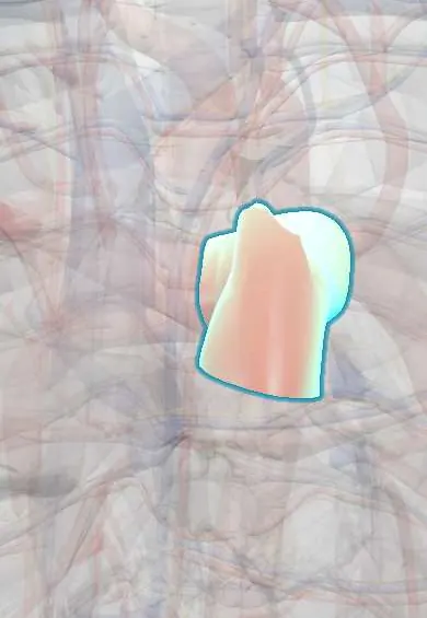
Pulmonary trunk !
The pulmonary trunk is the large artery leaving the right ventricle; it carries oxygen-poor blood toward both lungs before it divides.
Opens in the Durgical app, no sign-up needed.
What it is
The large artery leaving the right ventricle, splitting into the right and left pulmonary arteries.
What it does
Carries oxygen-poor blood to the lungs (an artery that carries 'blue' blood).
Key facts
- Origin
- Conus arteriosus of right ventricle via pulmonary valve, behind left 3rd costal cartilage
- Branches
- Right and left pulmonary arteries, dividing under aortic arch (about T5 level)
- Supplies
- Lungs: deoxygenated blood for gas exchange; in fetus, ductus arteriosus to aorta
Relations
- Medial
- Ascending aorta, around which it spirals; both share a sleeve of serous pericardium
- Lateral
- Left auricle, with the left coronary artery emerging between the two
- Behind
- The transverse pericardial sinus, which separates it and the aorta from the atria and the superior vena cava; at its end, the left main bronchus
- Above
- Concavity of the aortic arch, joined to the left pulmonary artery by the ligamentum arteriosum; the superficial cardiac plexus lies here
How to find it
The pulmonary valve projects behind the left 3rd costal cartilage near the sternum, and its sounds are best heard in the 2nd left intercostal space. On a frontal chest X-ray the trunk forms the bulge on the left heart border just below the aortic knob.
On imaging
On a frontal chest X-ray the left heart border shows, from top to bottom: aortic knob, pulmonary trunk, left atrial appendage and left ventricle. A prominent trunk suggests pulmonary hypertension or dilatation after pulmonary valve stenosis. CT and MRI measure its width against the ascending aorta.
Clinical notes
- A saddle embolus straddling the bifurcation can cause sudden right ventricular failure
- In cardiac surgery the transverse pericardial sinus behind the trunk and aorta is used to pass a clamp or tape around both
- Tetralogy of Fallot narrows the right ventricular outflow into the trunk; transposition of the great arteries places it on the left ventricle
Development
The truncus arteriosus is divided by the spiral aorticopulmonary septum, built with neural crest cells. Failure of septation leaves a persistent truncus arteriosus; a septum that does not spiral gives transposition of the great arteries.
Related conditions
- Transposition of the great arteries
- Pulmonary hypertension and cor pulmonale
- Pulmonary valve stenosis
- Truncus arteriosus, TAPVR and tricuspid atresia
- Pulmonary embolism
- Patent ductus arteriosus
- Tetralogy of Fallot
Test yourself
Think of the answer, then tap the question to see it.
Where is the pulmonary valve heard, and what does a loud pulmonary component of the second heart sound suggest?
In the 2nd left intercostal space beside the sternum; a loud pulmonary component suggests pulmonary hypertension.
On CT, what does a pulmonary trunk wider than the ascending aorta at the same level suggest?
Pulmonary hypertension.
Which pericardial space lies behind the pulmonary trunk and ascending aorta, and how is it used in surgery?
The transverse pericardial sinus, which separates them from the atria and superior vena cava; a clamp or tape is passed through it around both vessels.
What is a saddle embolus of the pulmonary trunk and what can it cause?
A clot straddling the bifurcation of the pulmonary trunk; it can cause sudden right ventricular failure.
Educational information, not medical advice. For any symptom, see a health professional.
Its names
- Official Latin (TA2)
- truncus pulmonalis TA2 4075
- Official Spanish (DeCS)
- Tronco pulmonar DeCS D011651
- English (TA2)
- Pulmonary trunk
- Also called
- arteria pulmonalis
Related
Sources: Terminologia Anatomica (TA2); DeCS; Anatomy, Thorax, Heart Great Vessels; Anatomy, Thorax, Lungs.
Spotted a mistake?We are always improving for the community. Tell us what is wrong and our team will review it.Report a mistake
Representative model, for educational purposes only.