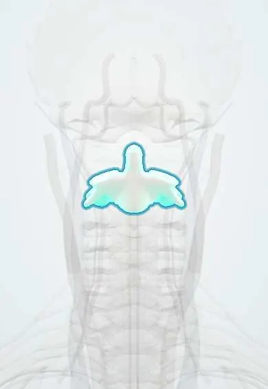
Axis (C2) !
The axis (C2) is the second neck vertebra; its tooth-like peg, the dens, is the pivot the head turns on to shake it "no".
Official name: Axis
Opens in the Durgical app, no sign-up needed.
What it is
The second neck vertebra, with a tooth-like peg (the dens) that sticks up through the ring of the atlas.
What it does
Acts as the pivot the head and atlas turn around when you shake your head 'no'.
Key facts
- Joints with
- Atlas above (median atlantoaxial joint at the dens, lateral atlantoaxial joints); C3 below (C2–C3 disc and facet joints)
- Landmarks
- Dens (odontoid process) rising from the body, held by transverse and alar ligaments; large bifid spinous process, first one felt below the occiput; transverse foramina
Relations
- Above
- The dens sits inside the ring of the atlas (C1), held against its anterior arch by the transverse ligament of the atlas; the alar ligaments tie its tip to the occipital condyles
- In front
- Body and dens behind the oropharynx, with longus colli and the prevertebral fascia between; swelling of this soft tissue on a lateral X-ray hints at a fracture
- Behind
- The large spinous process anchors rectus capitis posterior major and obliquus capitis inferior (suboccipital muscles), semispinalis cervicis and the nuchal ligament
- Lateral
- Transverse foramina with the vertebral artery, which bends outwards here toward the wider-set foramina of C1; the C2 ganglion lies behind the lateral atlantoaxial joint
- Contents
- Spinal cord with ample room; the tectorial membrane, the upward continuation of the posterior longitudinal ligament, covers the back of the body and dens on its way to the clivus
How to find it
With the head bent slightly forward, a finger slid down the midline from the external occipital protuberance drops into a soft hollow and then meets the first firm bony point: the broad, bifid spinous process of the axis (C2).
Examination
- Cervical flexion-rotation test: lying supine, the neck is fully flexed to lock the lower levels and the head is turned to each side; clearly less rotation to one side points to the C1–C2 segment, for example as a source of headache arising from the neck
On imaging
Open-mouth AP view for the dens: the incisors or the occiput can draw a false line across its base (Mach band). Lateral view: the dens, the ring outline over the C2 body (broken in type III fractures) and the posterior cervical line from C1 to C3, which separates a hangman's fracture from the normal C2–C3 pseudosubluxation of children. CT confirms; MRI shows ligaments and cord.
Common problems
- Dens (odontoid) fractures, common in older adults after a fall.
- 'Hangman's' fracture through the back arch, from sudden hyperextension.
- Arthritis of the C1-C2 joints: pain at the base of the skull when turning the head.
How it is diagnosed
CT for fractures; MRI for the ligaments and spinal cord; an open-mouth X-ray view shows the dens.
Treatment
- Collar or halo brace for many stable fractures.
- Surgery (a screw through the dens, or a C1-C2 fusion) for displaced or non-healing fractures.
Clinical notes
- Type II fractures are managed according to displacement, age and the risk of non-union: collar or halo, an anterior screw through the dens, or C1–C2 fusion, which gives up much of neck rotation
- Extension teardrop: a small fragment pulled off the front lower corner of C2 in older adults; usually stable, unlike the flexion teardrop of the lower neck
- Screws in the narrow C2 pars and pedicle are planned on CT, because the vertebral artery can run unusually high and medial in the body
- Erosion of the dens by rheumatoid pannus, or a dens that rises into the foramen magnum (cranial settling), can compress the brainstem
Anatomical variants
- Os odontoideum: a separate ossicle with smooth, rounded edges above a short dens; a congenital or post-traumatic origin is debated, and it can make C1–C2 unstable
- Persistent ossiculum terminale: the tip of the dens stays unfused, usually without consequence
- High-riding vertebral artery in the C2 body or pedicle
Development
The dens is in origin the body of the atlas, fused onto C2. In young children the cartilage plate between dens and body (subdental synchondrosis) is still open and can look like a type III fracture; it closes in early childhood (texts give roughly 3–7 years).
Related conditions
Test yourself
Think of the answer, then tap the question to see it.
Which type of dens fracture of the axis (C2) is the most common and the most prone to non-union?
Type II (Anderson–D'Alonzo), which crosses the base of the dens.
What is a hangman's fracture of the axis (C2)?
A traumatic spondylolisthesis of the axis: both pars interarticularis break in hyperextension with axial load; the canal widens, so the cord is often spared.
Which nerve comes from the dorsal ramus of the C2 nerve, and what symptom can it cause?
The greater occipital nerve, a source of occipital headache; the C2 nerve leaves behind the lateral atlantoaxial joint, not through a typical foramen.
What is the developmental origin of the dens of the axis (C2)?
It is the body of the atlas fused onto C2; in young children the open subdental synchondrosis can look like a type III fracture.
Educational information, not medical advice. For any symptom, see a health professional.
Its names
- Official Latin (TA2)
- axis TA2 1050
- Official Spanish (DeCS)
- Vértebra cervical axis DeCS D001368
- English (TA2)
- Axis
- Also called
- vertebra C2 · Axis, Cervical Vertebra · Odontoid Process · os axiale · axis bone · epistropheus
Related
- C2–C3 disc
- Vertebral artery
- Alar ligaments
- Anterior longitudinal ligament
- Atlas–axis joints
- Endplates
- Neck facet joints
- Interspinales
Sources: Terminologia Anatomica (TA2); DeCS; Anatomy, Head and Neck: Cervical Vertebrae; Anatomy, Head and Neck: Atlantoaxial Joint.
Spotted a mistake?We are always improving for the community. Tell us what is wrong and our team will review it.Report a mistake
Representative model, for educational purposes only.