Cardiovascular system · Head and neck
Basilar artery !
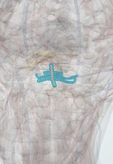
The basilar artery runs up the front of the brainstem and feeds the brainstem, cerebellum and back of the brain; a clot here is a severe stroke.
Opens in the Durgical app, no sign-up needed.
What it is
The single artery on the front of the pons, formed by the two vertebral arteries.
What it does
Feeds the brainstem, cerebellum and back of the brain; a clot here is a life-threatening stroke.
Key facts
- Origin
- Union of the two vertebral arteries at the pontomedullary junction
- Branches
- AICA; pontine; labyrinthine (usually from AICA); SCA; ends as the two posterior cerebral arteries (oculomotor nerve between SCA and PCA)
- Supplies
- Pons, cerebellum, inner ear, midbrain
Relations
- Course
- Formed at the lower border of the pons, it runs up in the basilar groove on the front of the pons within the prepontine cistern and divides at the upper border into the two posterior cerebral arteries
- In front
- Clivus and dorsum sellae, covered by dura
- Behind
- Pons
- Lateral
- Abducens nerves climbing forward from the pontomedullary junction; near the top, the oculomotor nerves between the superior cerebellar and posterior cerebral arteries
How to find it
It cannot be felt. It runs up the front of the pons in the pontine cistern, resting on the clivus, and is seen best on sagittal MRI or CT angiography; transcranial Doppler reaches it through the foramen magnum with the probe below the occiput and the head bent forward.
On imaging
CT can miss brainstem infarcts because of bone artefact; diffusion-weighted MRI is more sensitive. A hyperdense basilar sign on CT, and CT or MR angiography, show the occlusion.
Clinical notes
- The basilar tip is one of the classic aneurysm sites, often treated endovascularly
- Pontine haemorrhage, often from high blood pressure, gives coma, pinpoint pupils and weakness of all four limbs
- An elongated, widened (dolichoectatic) basilar artery can press on cranial nerves or the brainstem
Anatomical variants
- Fenestration (a short split segment), seen most often near its lower end
- A persistent trigeminal artery can join it to the internal carotid artery
Development
It forms by fusion of paired longitudinal neural arteries, which is why partial splits (fenestrations) occur.
Related conditions
Test yourself
Think of the answer, then tap the question to see it.
How is the basilar artery formed and how does it end?
By the union of the two vertebral arteries at the lower border of the pons; it ends by dividing into the two posterior cerebral arteries.
What is locked-in syndrome, and which artery is involved?
Infarction of the front of the pons from basilar artery occlusion: awake but unable to move or speak, except vertical eye movements and blinking.
What is the top of the basilar syndrome?
An embolus at the basilar tip affecting the midbrain, thalami and occipital lobes: drowsiness, eye movement problems, visual loss and memory problems.
Educational information, not medical advice. For any symptom, see a health professional.
Its names
- Official Latin (TA2)
- arteria basilaris TA2 4548
- Official Spanish (DeCS)
- Arteria Basilar DeCS D001488
- English (TA2)
- Basilar artery
- Also called
- basilar trunk · arteria basialis
Related
Sources: Terminologia Anatomica (TA2); DeCS; Anatomy, Head and Neck: Basilar Artery.
Spotted a mistake?We are always improving for the community. Tell us what is wrong and our team will review it.Report a mistake
Representative model, for educational purposes only.