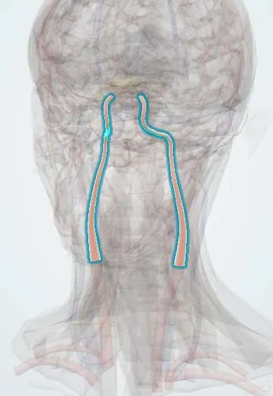
Cardiovascular system · Head and neck
Internal carotid artery !
The internal carotid artery climbs into the skull and feeds most of the brain and the eye; narrowing of it is a major cause of stroke.
Opens in the Durgical app, no sign-up needed.
What it is
The branch with no branches in the neck, entering the skull through the carotid canal.
What it does
Feeds most of the brain and the eye; narrowing here is a major cause of stroke.
Key facts
- Origin
- Carotid bifurcation
- Branches
- None in the neck; ophthalmic, posterior communicating, anterior choroidal, ACA, MCA
- Supplies
- Most of the brain and the eye; enters through the carotid canal, crosses the cavernous sinus
Relations
- Course
- Rises in the carotid sheath in front of the upper cervical transverse processes, enters the carotid canal of the petrous temporal bone, turns forward above the foramen lacerum, makes an S-bend (the carotid siphon) in the cavernous sinus, pierces the dura medial to the anterior clinoid process and divides beside the optic chiasm into the anterior and middle cerebral arteries
- Lateral
- Internal jugular vein in the neck, with the vagus nerve behind and between them; in the cavernous sinus, the abducens nerve, and in the sinus wall the oculomotor, trochlear, ophthalmic and maxillary nerves
- Medial
- Wall of the pharynx and, higher up, the region of the palatine tonsil; inside the skull, the sphenoid sinus and the pituitary gland in the sella turcica
- Behind
- Longus capitis, the sympathetic trunk and the superior cervical ganglion, whose fibres form a plexus on the artery and travel with it into the skull
- Superficial
- Sternocleidomastoid; the hypoglossal nerve and the posterior belly of the digastric cross it; the styloid process and its muscles separate it from the external carotid artery
How to find it
The carotid pulse is felt at the front border of the sternocleidomastoid level with the top edge of the thyroid cartilage, where the common carotid usually divides (about C3–C4). The internal carotid begins behind and lateral to the external and gives no branches in the neck.
Examination
- Listening over the bifurcation for a bruit: it may signal narrowing, but its absence does not rule out severe stenosis, so duplex ultrasound is the usual next step
On imaging
Duplex ultrasound: usually posterolateral, with a wider bulb, no neck branches and a low-resistance waveform with flow through diastole; peak velocities grade a stenosis. CT or MR angiography shows the petrous, cavernous and intracranial parts and the siphon.
Common problems
- Carotid stenosis: plaque narrows the artery near the bifurcation; pieces can break off and cause a stroke or a mini-stroke (TIA).
- Brief loss of vision in one eye ('a curtain') can be a warning sign.
How it is diagnosed
Listening for a bruit, Doppler ultrasound, CT or MR angiography.
Treatment
- Antiplatelets, statins and blood pressure control.
- Carotid endarterectomy: the plaque is removed through a neck incision.
- Carotid stenting through a catheter in some patients.
Clinical notes
- Stenosis near its origin sheds emboli; endarterectomy or stenting is chosen by symptoms and degree of narrowing, and during clamping the brain depends on the circle of Willis
- TIA and amaurosis fugax are warnings: the risk of stroke is highest in the first days, so they are assessed urgently
- Carotid–cavernous fistula: a pulsating, bulging, red eye with a bruit over the orbit, after head trauma or from a ruptured cavernous aneurysm
- A tortuous internal carotid can lie close to the tonsil bed or bulge into the back of the pharynx, a hazard in tonsil surgery or drainage of a peritonsillar abscess
- In the cavernous sinus it grooves the sphenoid body and often bulges into the lateral wall of the sphenoid sinus, where the bone can be thin or missing: a hazard in endoscopic sinus and transsphenoidal pituitary surgery
Anatomical variants
- Kinking, coiling and tortuosity of the cervical part are common, especially in older people
- A persistent trigeminal artery, a fetal link between the cavernous internal carotid and the basilar artery, is the commonest persistent carotid–basilar anastomosis, though still rare
Development
The first part comes from the third pharyngeal arch artery and the rest from the cranial part of the dorsal aorta.
Related conditions
- Ischaemic stroke
- Transient ischaemic attack
- Carotid artery stenosis
- Atherosclerosis and dyslipidaemia
- Horner syndrome
Test yourself
Think of the answer, then tap the question to see it.
What branches does the internal carotid artery give in the neck?
None; inside the skull it gives the ophthalmic, posterior communicating and anterior choroidal arteries, then the ACA and MCA.
What is amaurosis fugax and how does it relate to the internal carotid artery?
A painless curtain over one eye for minutes: a retinal TIA from emboli entering the ophthalmic artery, its first intracranial branch.
How does internal carotid artery dissection typically present?
Neck, face or head pain with a partial Horner syndrome (ptosis and miosis, sweating spared); a stroke can follow hours to days later.
Which cranial nerve is often the first affected by a cavernous internal carotid aneurysm, and why?
The abducens nerve (VI): it lies beside the artery inside the cavernous sinus.
What are the signs of a carotid–cavernous fistula?
A pulsating, bulging, red eye with a bruit over the orbit, after head trauma or a ruptured cavernous aneurysm.
Educational information, not medical advice. For any symptom, see a health professional.
Its names
- Official Latin (TA2)
- carotis interna TA2 4463
- Official Spanish (DeCS)
- Arteria Carótida Interna DeCS D002343
- English (TA2)
- Internal carotid artery
Related
Sources: Terminologia Anatomica (TA2); DeCS; Anatomy, Head and Neck: Internal Carotid Arteries; Anatomy of the Human Body (20th ed.): Surface Markings of Special Regions of the Head and Neck (Lea & Febiger (Gray's Anatomy, 1918), 1918) · adapted, Public domain; 20.2 Blood Flow, Blood Pressure, and Resistance (OpenStax (Rice University), 2022) · adapted, CC BY 4.0; Tratado de anatomía humana (9.ª ed.), tomo II, Libro IV, sección 2 Arterias, caps. I-III (sistema de la arteria pulmonar; aorta y ramas del cayado hasta la carótida interna).
Spotted a mistake?We are always improving for the community. Tell us what is wrong and our team will review it.Report a mistake
Representative model, for educational purposes only.