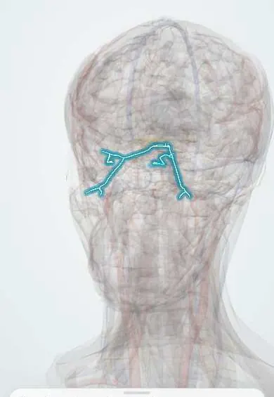
Nervous system · Head and neck
Oculomotor nerve (III) !
The oculomotor nerve (cranial nerve III) moves most eye muscles, lifts the eyelid and narrows the pupil; if compressed, the lid droops and the pupil widens.
Official name: Oculomotor nerve
Opens in the Durgical app, no sign-up needed.
What it is
The nerve from the midbrain to most eye muscles, the eyelid lifter and the pupil.
What it does
Moves the eye up, down and in, lifts the lid and narrows the pupil; when compressed the eye turns down and out with a wide pupil.
Key facts
- Roots
- Nuclei: oculomotor and Edinger-Westphal (midbrain); exits the interpeduncular fossa; superior orbital fissure
- Moves
- Superior division: SR, levator palpebrae; inferior division: MR, IR, IO and the parasympathetic root to the ciliary ganglion (from the nerve to IO): sphincter pupillae, ciliary muscle
- Feeling from
- None
Relations
- Course
- Leaves the midbrain in the interpeduncular fossa, passes between the posterior cerebral artery and the superior cerebellar artery, runs forward beside the posterior communicating artery, through the roof and lateral wall of the cavernous sinus and into the orbit through the superior orbital fissure, inside the common tendinous ring, where it splits into superior and inferior divisions
- Above
- It is the highest nerve in the lateral wall of the cavernous sinus, above the trochlear nerve (IV) and the V1 ophthalmic nerve
- Lateral
- At the tentorial notch, the uncus of the temporal lobe and the free edge of the tentorium; a swelling hemisphere pushes the uncus onto the nerve
How to find it
It cannot be felt; it is assessed through eye movements, the eyelid and the pupil. On MRI or in dissection it is found emerging between the cerebral peduncles and running forward between the posterior cerebral and superior cerebellar arteries.
Examination
- Eye movements in an H pattern: weak adduction, elevation and depression point to CN III; in primary gaze the eye rests down and out
- Pupillary light reflex: a dilated pupil that does not constrict to direct or consensual light on that side means the efferent limb (CN III) is affected
- Near (accommodation) reflex: on the affected side the pupil does not constrict and the eye does not converge
- Eyelid: ptosis from levator palpebrae weakness, often complete in a full palsy
On imaging
When the pupil is involved, CT or MR angiography looks for a posterior communicating artery aneurysm; MRI shows the midbrain nucleus, the cisternal segment and the cavernous sinus.
Common problems
- Palsy: droopy lid, the eye turned down and out, double vision.
- A palsy with a wide pupil can mean an aneurysm pressing on it: an emergency.
- Diabetes can cause a palsy that usually spares the pupil.
How it is diagnosed
Eye and pupil exam; urgent CT/MR angiography when the pupil is involved.
Treatment
- Treat the cause.
- Securing an aneurysm (clip or coils).
- Most diabetic palsies recover within months.
Clinical notes
- Weber syndrome: a lesion of the midbrain gives a CN III palsy on the same side with weakness of the opposite side of the body
- Cavernous sinus disease (thrombosis, tumour, carotid-cavernous fistula) can affect III together with IV, V1, V2 and VI
- The ptosis of a CN III palsy is usually marked and the pupil large, unlike the mild ptosis with a small pupil of Horner syndrome
Memory aid
Eye muscle nerves: "LR6 SO4, the rest 3": lateral rectus by VI, superior oblique by IV, all the others by III.
Related conditions
- Oculomotor (third) nerve palsy
- Subarachnoid haemorrhage
- Traumatic brain injury
- Epidural haematoma
- Pituitary adenoma
Test yourself
Think of the answer, then tap the question to see it.
What is the eye position in a complete oculomotor nerve (III) palsy, and why?
Down and out, with marked ptosis and a fixed dilated pupil: the lateral rectus (VI) and superior oblique (IV) act unopposed.
In an oculomotor nerve palsy, what does pupil involvement suggest compared with pupil sparing?
Pupil involved: compression, classically a posterior communicating artery aneurysm (emergency). Spared: usually diabetic or hypertensive microvascular palsy.
Why does outside compression of the oculomotor nerve affect the pupil first?
The parasympathetic pupil fibres run on the surface of the nerve, while small-vessel ischaemia damages its core.
What early sign does uncal herniation produce through the oculomotor nerve?
A fixed, dilated pupil on the side of the mass, as the uncus presses the nerve against the tentorium.
What is Weber syndrome?
A midbrain lesion giving an oculomotor nerve (III) palsy on the same side with weakness of the opposite side of the body.
Educational information, not medical advice. For any symptom, see a health professional.
Its names
- Official Latin (TA2)
- nervus oculomotorius TA2 6187
- Official Spanish (DeCS)
- Nervio Oculomotor DeCS D009802
- English (TA2)
- Oculomotor nerve
- Also called
- cranial nerve III
Related
- Cavernous sinus
- Posterior communicating artery
- Superior rectus
- Inferior rectus
- Medial rectus
- Inferior oblique
- Levator palpebrae
- Iris
Sources: Terminologia Anatomica (TA2); DeCS; Neuroanatomy, Cranial Nerve 3 (Oculomotor).
Spotted a mistake?We are always improving for the community. Tell us what is wrong and our team will review it.Report a mistake
Representative model, for educational purposes only.