Nervous system · Head and neck
Cerebellum !
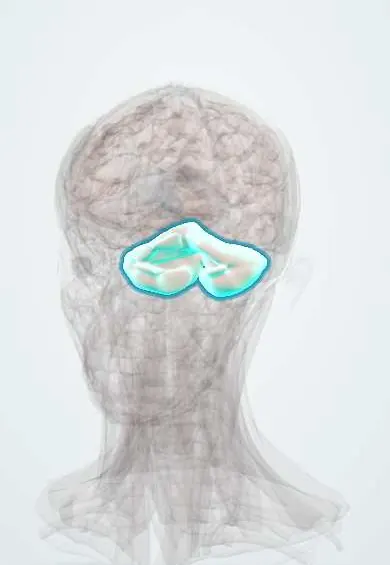
The cerebellum is the 'little brain' at the back of the head below the cerebrum; it coordinates movement, balance and timing, and damage makes movement clumsy.
Official name: Anterior quadrangular lobule
Opens in the Durgical app, no sign-up needed.
What it is
The 'little brain' at the back below the cerebrum, with finely folded hemispheres.
What it does
Coordinates movement, balance and timing; damage makes movements clumsy (ataxia).
Key facts
- Where
- Posterior cranial fossa under the tentorium
- Blood supply
- SCA, AICA, PICA
- Function
- Coordination, balance, motor learning (same side of the body)
Relations
- Above
- Tentorium cerebelli, and above it the occipital lobes
- In front
- Fourth ventricle, pons and medulla oblongata, joined to them by the three cerebellar peduncles
- Lateral
- Sigmoid sinus and the back of the petrous temporal bone; the facial and vestibulocochlear nerves at the cerebellopontine angle
- Behind
- Occipital bone of the posterior cranial fossa, with the transverse sinuses and the confluence of sinuses
- Below
- Cerebellar tonsils above the foramen magnum and the cisterna magna
How to find it
Fills the posterior fossa below the tentorium, behind the pons and medulla, roofing the fourth ventricle. On the skull its upper limit follows the transverse sinus from the inion toward the mastoid (superior nuchal line), the landmark for suboccipital openings. Its tonsils lie beside the medulla above the foramen magnum; a tonsil tip over about 5 mm below it on sagittal MRI suggests Chiari I.
Examination
- Finger-to-nose: the finger overshoots (dysmetria) and shakes more as it nears the target (intention tremor)
- Heel-to-shin: the heel wavers off the shin; mainly tests the legs and the anterior vermis
- Rapid alternating movements (patting the hand palm up, palm down): slow, irregular rhythm is dysdiadochokinesia
- Tandem gait: falling to one side on heel-to-toe walking; Romberg test is usually negative, as the unsteadiness is present with the eyes open
On imaging
MRI is the study of choice; on CT the posterior fossa is blurred by streak artefact from the dense skull base. A swollen cerebellar infarct can be seen compressing the fourth ventricle.
Common problems
- Cerebellar stroke or bleeding: sudden dizziness, vomiting and inability to walk.
- Alcohol and some medicines impair it: unsteady walking, slurred speech.
How it is diagnosed
Coordination tests (finger-to-nose, heel-to-shin); CT or MRI.
Treatment
- Treat the cause; surgery to relieve pressure for large strokes or bleeds; balance rehabilitation.
Clinical notes
- Medulloblastoma, the most common malignant brain tumour in children, arises in the midline cerebellum and can spread through the CSF
- Pilocytic astrocytoma of childhood typically forms a cyst with a nodule in a cerebellar hemisphere; haemangioblastoma is the adult cerebellar tumour linked to von Hippel–Lindau disease
- A large cerebellar infarct or bleed can swell, block the fourth ventricle and compress the brainstem; suboccipital decompression may be lifesaving
- Otogenic cerebellar abscess: middle ear and mastoid infection can spread through the thin posterior surface of the petrous bone or along a thrombosed sigmoid sinus into the adjacent cerebellar hemisphere
Development
Grows from the dorsal part of the metencephalon (the rhombic lips) and keeps developing after birth. Chiari II and Dandy–Walker malformations are hindbrain anomalies that often come with hydrocephalus.
Memory aid
Cerebellar signs: DANISH (Dysdiadochokinesia, Ataxia, Nystagmus, Intention tremor, Scanning speech, Hypotonia). Deep nuclei, lateral to medial: 'Don't Eat Greasy Food'.
Related conditions
- Maple syrup urine disease
- Congenital disorder of glycosylation type Ia (PMM2-CDG)
- Sandhoff disease
- Ataxia-telangiectasia
- Prion diseases (Creutzfeldt–Jakob)
- Mercury poisoning
- Menkes disease
- Intracerebral haemorrhage
- Acute alcohol (ethanol) intoxication
- Anticonvulsant toxicity (phenytoin, carbamazepine, valproate)
- Rabies
- Essential tremor
- Brain metastases
- Childhood brain tumours (medulloblastoma, pilocytic astrocytoma, craniopharyngioma)
- Friedreich ataxia
- Multiple system atrophy (MSA)
- Spinocerebellar ataxias (autosomal dominant ataxias)
- Alcohol use disorder
- Autism spectrum disorder (ASD)
- Cannabis use disorder
- Lithium toxicity
Test yourself
Think of the answer, then tap the question to see it.
On which side of the body do cerebellar signs appear?
On the same side as the lesion, because its output crosses in the midbrain and the corticospinal tract crosses back.
What are the classic cerebellar signs?
Dysmetria, intention tremor, dysdiadochokinesia, nystagmus, scanning speech, hypotonia and a broad-based unsteady gait.
What does the finger-to-nose test show in cerebellar disease?
The finger overshoots (dysmetria) and shakes more as it nears the target (intention tremor).
Why is the Romberg test usually negative in cerebellar ataxia?
The unsteadiness is present with the eyes open too, so closing them adds little.
Which is the most common malignant brain tumour in children, and where does it arise?
Medulloblastoma, in the midline cerebellum; it can spread through the CSF.
Educational information, not medical advice. For any symptom, see a health professional.
Its names
- Official Latin (TA2)
- lobulus quadrangularis anterior TA2 5808
- Official Spanish (DeCS)
- Cerebelo DeCS D002531
- English (TA2)
- Anterior quadrangular lobule
Related
Sources: Terminologia Anatomica (TA2); DeCS; Neuroanatomy, Cerebellum; 13.1 The Embryologic Perspective (OpenStax (Rice University), 2022) · adapted, CC BY 4.0; 13.2 The Central Nervous System (OpenStax (Rice University), 2022) · adapted, CC BY 4.0; Tratado de anatomía humana (9.ª ed.), tomo II, Libro V, sección 2 (Encéfalo), capítulos I-V: bulbo, protuberancia, cerebelo, cuarto ventrículo, mesencéfalo.
Spotted a mistake?We are always improving for the community. Tell us what is wrong and our team will review it.Report a mistake
Representative model, for educational purposes only.