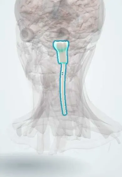
Brainstem stroke syndromes (Wallenberg and others) !
Strokes in the midbrain, pons or medulla. They cause "crossed" signs: cranial nerve problems on one side and body weakness or numbness on the other.
Updated 2026-11-01
Structures involved
Tap one to see what it is and open it in 3D.
- Medulla oblongata
- Pons
- Midbrain
- Vertebral artery
- Basilar artery
- Cerebellar arteries
- Posterior cerebral artery
- Cerebellum
- Cerebellar peduncles
- Thalamus
- Oculomotor nerve (III)
- Trigeminal nerve (V)
- Abducens nerve (VI)
- Facial nerve (VII)
- Vestibulocochlear nerve (VIII)
- Vagus nerve
- Hypoglossal nerve (XII)
Why it happens
- Infarction: red vs white, and what decides the damage
- Systemic (arterial) thromboembolism
- Somatosensory pathways: dorsal column and anterolateral
What it is
A group of stroke syndromes caused by occlusion of small branches of the vertebral and basilar arteries (posterior circulation). The classic pattern is ipsilateral cranial nerve deficits with contralateral long-tract (motor or sensory) signs, which lets the level and side of the lesion be localized.
Who gets it
Posterior circulation strokes are about one in five ischemic strokes. Lateral medullary (Wallenberg) syndrome is the most common brainstem syndrome. In Colombia and Latin America the main drivers are the same as for any ischemic stroke: hypertension, diabetes, smoking and atrial fibrillation; vertebral artery dissection is an important cause in young adults.
Causes
- Atherosclerosis or thrombosis of the vertebral or basilar artery
- Small-vessel (lacunar) disease of paramedian penetrating branches
- Cardioembolism (e.g. atrial fibrillation, Chagas cardiomyopathy)
- Vertebral artery dissection after neck trauma or sudden neck movements
Risk factors
- Hypertension
- Diabetes mellitus
- Smoking and dyslipidemia
- Atrial fibrillation and other cardiac sources of emboli
- Older age; neck manipulation or trauma in younger patients
Pathophysiology
The brainstem is packed with cranial nerve nuclei and with long tracts that cross at different levels. Medial (paramedian) branches supply the corticospinal tract, medial lemniscus and the nuclei of nerves III, VI and XII; lateral (circumferential) branches such as the posterior inferior cerebellar artery (PICA) and anterior inferior cerebellar artery (AICA) supply the spinothalamic tract, the spinal trigeminal nucleus, vestibular nuclei, nucleus ambiguus and sympathetic fibres. A small infarct therefore produces a characteristic combination of signs, and the "rule of 4" (medial versus lateral structures, and cranial nerves III-IV midbrain, V-VIII pons, IX-XII medulla) helps localize it.
Classification
- Lateral medullary (Wallenberg) syndrome: PICA or vertebral artery; ipsilateral facial pain and temperature loss, contralateral body pain and temperature loss, dysphagia and hoarseness (nucleus ambiguus), vertigo, nystagmus, ipsilateral ataxia and Horner syndrome
- Medial medullary syndrome: anterior spinal artery or vertebral branches; contralateral hemiparesis and loss of position sense, ipsilateral tongue deviation (nerve XII)
- Lateral pontine syndrome: AICA; facial paralysis, hearing loss, vertigo and ataxia on the same side, contralateral pain and temperature loss
- Medial pontine syndrome: paramedian basilar branches; contralateral hemiparesis, ipsilateral abducens palsy
- Weber syndrome (midbrain): ipsilateral oculomotor palsy with contralateral hemiparesis
- Locked-in syndrome: bilateral ventral pons (basilar artery); quadriplegia and loss of speech with preserved consciousness and vertical eye movements
Symptoms and signs
- Sudden vertigo, nausea and vomiting
- Double vision and abnormal eye movements or nystagmus
- Difficulty swallowing, hoarse voice and slurred speech
- Unsteady gait or clumsy limbs (ataxia)
- Facial numbness or weakness on one side with body numbness or weakness on the opposite side
- Drooping eyelid and small pupil (Horner syndrome) in lateral medullary infarcts
- Hiccups that do not stop in some lateral medullary strokes
Diagnosis
- Clinical
- Sudden onset of crossed findings localizes the lesion. Bedside neurological examination with the HINTS test (head impulse, nystagmus, test of skew) helps separate central from peripheral vertigo; a swallowing screen is done before anything is given by mouth. The NIHSS underestimates posterior strokes.
- Tests
- Capillary glucose at once, blood count, coagulation, electrolytes, renal function and lipids. ECG and cardiac rhythm monitoring look for atrial fibrillation; echocardiography looks for embolic sources.
- Imaging
- Non-contrast CT first excludes hemorrhage but often misses posterior fossa infarcts. Diffusion-weighted MRI is the most sensitive test. CT or MR angiography shows vertebral or basilar occlusion and dissection.
Differential diagnosis
- Peripheral vestibular disorders (vestibular neuritis, benign paroxysmal positional vertigo)
- Cerebellar hemorrhage or infarct
- Multiple sclerosis with a brainstem plaque
- Myasthenia gravis or Guillain-Barré syndrome (Miller Fisher variant)
- Posterior fossa tumour
- Wernicke encephalopathy
Goal of treatment
Restore blood flow as fast as possible when eligible, protect the airway and prevent aspiration, prevent edema and herniation in large infarcts, and start secondary prevention according to the cause.
Treatment
- Acute
- Intravenous thrombolysis (alteplase or tenecteplase) within the time window if there are no contraindications; mechanical thrombectomy for basilar artery occlusion in selected patients.
- Medical
- Antiplatelet therapy after hemorrhage is excluded, high-intensity statins, and control of blood pressure and glucose. Anticoagulation for atrial fibrillation; antithrombotic treatment for vertebral dissection.
- Follow-up
- Nursing priorities: nothing by mouth until a formal swallowing assessment, head-of-bed elevation, frequent neurological checks for loss of consciousness, airway and oxygen saturation, and fall prevention because of vertigo and ataxia.
- Surgical
- Suboccipital decompressive craniectomy for large cerebellar infarcts with swelling, and ventricular drainage if hydrocephalus develops.
- Rehabilitation
- Early rehabilitation with physiotherapy, speech and swallowing therapy and vestibular exercises.
Complications
- Aspiration pneumonia
- Obstructive hydrocephalus and brainstem compression from cerebellar edema
- Locked-in syndrome or death with basilar occlusion
- Central respiratory and autonomic instability
- Persistent pain on the affected side (central post-stroke pain)
Prognosis
Many patients with lateral medullary syndrome recover well over weeks to months, although swallowing problems and pain may persist. Basilar artery occlusion has a high mortality and disability rate unless blood flow is restored early.
Test yourself
Think of the answer, then tap the question to see it.
Which artery is classically occluded in lateral medullary (Wallenberg) syndrome?
The posterior inferior cerebellar artery (PICA) or the intracranial vertebral artery.
Why does Wallenberg syndrome cause dysphagia and hoarseness?
The infarct damages the nucleus ambiguus, which supplies the muscles of the pharynx and larynx through nerves IX and X.
What is the pattern of pain and temperature loss in lateral medullary syndrome?
Ipsilateral face (spinal trigeminal nucleus) and contralateral body (spinothalamic tract).
A patient has a left oculomotor palsy and right hemiparesis. Where is the lesion?
Left midbrain (Weber syndrome).
Which imaging test is most sensitive for an acute brainstem infarct?
Diffusion-weighted MRI; non-contrast CT often misses small posterior fossa infarcts.
Related conditions
- Ischaemic stroke
- Transient ischaemic attack
- Intracerebral haemorrhage
- Horner syndrome
- Vestibular neuritis and labyrinthitis
- Benign paroxysmal positional vertigo (BPPV)
- Oropharyngeal dysphagia in older adults
- Atrial fibrillation
- Arterial hypertension
- Vestibular schwannoma (acoustic neuroma)
- Multiple sclerosis
Educational information, not medical advice. For any symptom, see a health professional.
Sources: Ischemic Stroke (MSD Manual Professional Edition, 2026).
Spotted a mistake?We are always improving for the community. Tell us what is wrong and our team will review it.Report a mistake
Representative model, for educational purposes only.