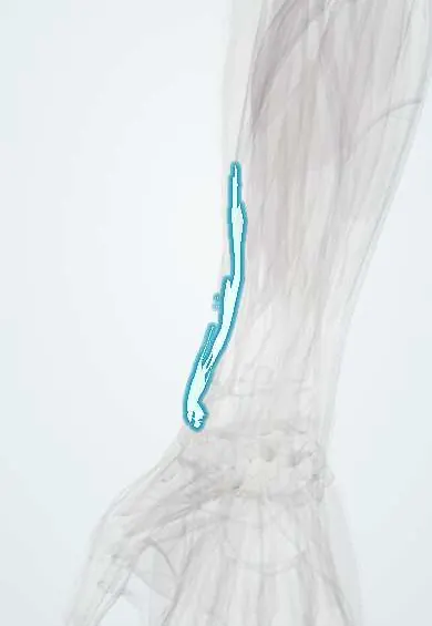
Brachioradialis tendon !
The brachioradialis tendon attaches the brachioradialis to the lower radius near the wrist; its pull can displace a distal radius fracture.
Opens in the Durgical app, no sign-up needed.
What it is
The flat tendon of brachioradialis ending on the radial styloid.
What it does
Passes the pull of brachioradialis to the radius; it can pull on a distal radius fracture.
Key facts
- Attaches
- Lateral side of the base of the radial styloid process
- Function
- Elbow flexion, strongest with the forearm mid-prone; tapped for the brachioradialis reflex (C6); the superficial radial nerve comes out from under it in the distal forearm
- Tight in
- Elbow extension
Relations
- Course
- The belly ends in mid-forearm in a flat tendon that runs down the lateral radius, crossed near the wrist by the APL and EPB, to the base of the radial styloid
- Deep
- Superficial radial nerve until it emerges; at its medial edge the radial artery; distally the radius
- Medial
- Radial artery and FCR tendon near the wrist
- Behind
- ECRL tendon, with the superficial radial nerve coming out between the two
- Superficial
- APL and EPB crossing over its end near the radial styloid; the lateral antebrachial cutaneous nerve and the cephalic vein on the fascia
How to find it
With the forearm halfway between pronation and supination and the elbow flexed against resistance, the muscle bulges on the outer forearm; its tendon is followed to the radial styloid, where it is tapped for the reflex a little above the tip.
Examination
- Brachioradialis reflex: the tendon is tapped just above the radial styloid with the forearm mid-prone; elbow flexion is normal, loss suggests a C5–C6 root or radial nerve problem, finger flexion alone is the inverted reflex
- Resisted elbow flexion with the forearm mid-prone makes it stand out; loss with an intact biceps points to a radial nerve lesion above the elbow
On imaging
Ultrasound and MRI show the tendon running along the radial side of the forearm to its insertion just above the radial styloid, with the superficial radial nerve emerging beside it. Radiographs do not show it, but its pull explains the shortening and loss of radial inclination seen on the PA view of a distal radius fracture.
Clinical notes
- Wartenberg syndrome: tight watch straps, casts or the scissoring of its tendon against the ECRL in pronation compress the superficial radial nerve, with numbness over the back of the thumb
- Brachioradialis to FPL transfer restores key pinch in tetraplegia
- In the volar approach to the radial styloid its tendon is released or lengthened to reduce the fracture
Anatomical variants
- A rare double belly with two superficial radial nerves is described
Development
Like the other arm and forearm muscles it forms in place from the mesoderm of the limb bud, not from somites; it belongs to the dorsal (extensor) muscle mass, so the radial nerve supplies it even though it flexes the elbow.
Test yourself
Think of the answer, then tap the question to see it.
Where does the brachioradialis tendon insert, and when is the muscle strongest?
On the lateral base of the radial styloid; it flexes the elbow, strongest with the forearm mid-prone.
Which reflex is tested at the brachioradialis tendon, and what does an inverted response suggest?
The brachioradialis reflex (C6); finger flexion without elbow flexion suggests spinal cord compression at C5–C6.
How does the brachioradialis tendon act in distal radius fractures?
As a deforming force: it pulls the styloid, shortening the fracture and tilting the fragment; it may be released during volar plating.
What is Wartenberg syndrome?
Compression of the superficial radial nerve where it emerges between the brachioradialis tendon and ECRL, causing numbness over the back of the hand.
Educational information, not medical advice. For any symptom, see a health professional.
Its names
- Official Latin (TA2)
- tendo musculi brachioradialis TA2 2496
- Official Spanish (DeCS)
- Tendón del músculo braquiorradial
- English (TA2)
- Brachioradialis tendon
Related
Sources: Terminologia Anatomica (TA2); Anatomy, Shoulder and Upper Limb, Forearm Brachioradialis Muscle; Anatomy of the Human Body (20th ed.): Development of the Muscles (Lea & Febiger (Gray's Anatomy, 1918), 1918) · adapted, Public domain.
Spotted a mistake?We are always improving for the community. Tell us what is wrong and our team will review it.Report a mistake
Representative model, for educational purposes only.