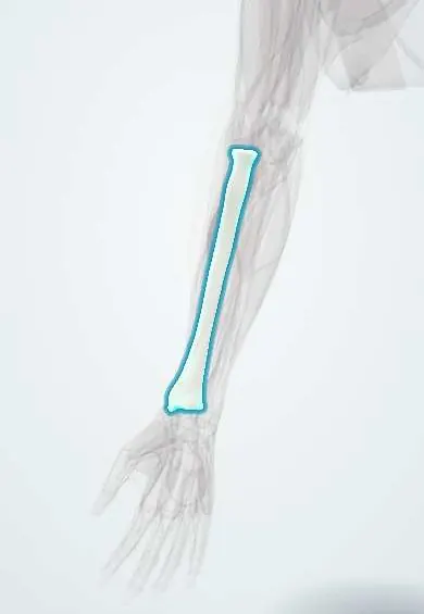
Radius !
The radius is the forearm bone on the thumb side; it turns around the ulna to flip the palm and carries most of the wrist's load.
Opens in the Durgical app, no sign-up needed.
What it is
The forearm bone on the thumb side; it widens at the wrist.
What it does
Rolls around the ulna to turn the palm up and down and carries most of the wrist's load.
Key facts
- Joints with
- Humerus (capitulum), ulna (proximal and distal radio-ulnar joints), scaphoid and lunate
- Landmarks
- Head, neck, radial tuberosity, interosseous border, ulnar notch, dorsal (Lister's) tubercle, styloid process
Relations
- Above
- Radial head against the capitulum, encircled by the annular ligament; the radial nerve divides in front of the elbow into the posterior interosseous nerve and the superficial radial nerve
- In front
- Distal biceps tendon to the radial tuberosity, with the bicipitoradial bursa; the supinator wraps the neck with the posterior interosseous nerve passing through it; FPL and pronator quadratus on the front of the shaft; the radial artery lies on the distal radius
- Behind
- Lister's tubercle separates the second extensor compartment (ECRL, ECRB) from the third (EPL), whose tendon turns around it; APL and EPB cross the back of the distal third
- Medial
- Ulna, joined by the interosseous membrane; the ulnar notch forms the distal radio-ulnar joint, stabilised by the TFCC
- Lateral
- Brachioradialis to the base of the styloid process; the first compartment tendons (APL, EPB) over the styloid; the superficial radial nerve crosses it under the skin; pronator teres inserts on the rough middle of the lateral surface, below the supinator, which wraps the upper third
How to find it
The radial head is felt just distal to the lateral epicondyle, turning under the thumb in pronation and supination. At the wrist, Lister's tubercle is felt on the back, in line with the index–middle finger cleft, and the styloid process on the thumb side, about 1 cm more distal than the ulnar styloid; a displaced distal radius fracture brings the two level.
On imaging
PA and lateral wrist views measure inclination, height and palmar tilt; a step of about 2 mm or more in the joint surface is significant, and CT maps intra-articular fractures. At the elbow, the radiocapitellar line (through the radial neck and head) should cross the capitulum on every view; if it does not, the radial head is dislocated.
Common problems
- Distal radius fracture (Colles fracture): the most common arm fracture, usually from a fall on an outstretched hand.
- Radial head fracture at the elbow, from a fall.
- A step in the wrist joint surface after a fracture can lead to arthritis.
How it is diagnosed
X-rays measure the tilt, length and joint surface; CT if the joint is involved. Check of the median nerve and the circulation.
Treatment
- Closed reduction (straightening without an incision) and a cast for stable fractures.
- Surgery with a plate and screws for unstable or displaced fractures.
- Hand therapy afterward to regain motion and grip.
Clinical notes
- The EPL tendon can rupture weeks after a distal radius fracture, typically an undisplaced one, where it turns around Lister's tubercle
- Acute carpal tunnel syndrome after a displaced distal radius fracture: the median nerve is checked before and after reduction
- Pulled elbow in toddlers: the radial head slips partly out of the annular ligament after a sudden pull on the hand
- The superficial radial nerve is at risk with wrist splints, casts and approaches to the radial styloid
Anatomical variants
- Congenital radio-ulnar synostosis, usually proximal, which fixes the forearm in pronation
- Radial longitudinal deficiency (hypoplastic or absent radius, radial club hand), which can occur with VACTERL association and other syndromes
Development
The radial head is the second elbow centre to appear (around age 3–5, the R of CRITOE). The distal physis provides most of the radius's growth in length, and Salter–Harris type II injuries of it are among the most common physeal fractures in children.
Memory aid
"MUGR": Monteggia = Ulna fracture, Galeazzi = Radius fracture; in each, the joint at the other end of the forearm is dislocated.
Related conditions
- Fanconi anaemia
- Osteoporosis
- Complex regional pain syndrome
- Hypertrophic osteoarthropathy and digital clubbing
- Forearm fractures (both-bone, Monteggia, Galeazzi)
- Distal radius fracture (Colles)
- Radial head subluxation (pulled elbow)
Test yourself
Think of the answer, then tap the question to see it.
How do Colles and Smith fractures of the distal radius differ?
Colles has dorsal tilt of the fragment (dinner-fork deformity); Smith has palmar tilt (garden-spade deformity).
What is a Galeazzi fracture of the radius?
A fracture of the distal third of the radial shaft with disruption of the distal radio-ulnar joint.
What are the usual normal X-ray values for the distal radius?
Radial inclination about 22°, radial height about 11 mm and palmar tilt about 11°.
Which tendon can rupture weeks after a distal radius fracture, and where?
The extensor pollicis longus tendon, where it turns around Lister's tubercle, typically after an undisplaced fracture.
What is the Essex-Lopresti injury of the radius?
A radial head fracture with a tear of the interosseous membrane and distal radio-ulnar joint injury, letting the radius migrate proximally.
Educational information, not medical advice. For any symptom, see a health professional.
Its names
- Official Latin (TA2)
- radius TA2 1210
- Official Spanish (DeCS)
- Radio
- English (TA2)
- Radius
Related
- EPL tendon
- Radial artery
- Interosseous arteries
- Annular ligament
- APL
- APL and EPB tendons
- Distal biceps tendon
- Bicipitoradial bursa
Sources: Terminologia Anatomica (TA2); Anatomy, Shoulder and Upper Limb, Forearm Radius; Anatomy, Shoulder and Upper Limb, Forearm Bones; Anatomía humana (4.ª ed.), tomo 1, cap. 54: Huesos del miembro superior.
Spotted a mistake?We are always improving for the community. Tell us what is wrong and our team will review it.Report a mistake
Representative model, for educational purposes only.