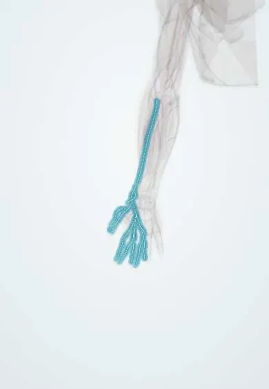
Superficial radial nerve !
The superficial radial nerve is the skin branch of the radial nerve; it gives feeling to the back of the hand on the thumb side.
Official name: Superficial branch of the radial nerve
Opens in the Durgical app, no sign-up needed.
What it is
A skin nerve that runs under brachioradialis and comes out on the thumb side of the wrist.
What it does
Feeling on the back of the thumb side of the hand.
Key facts
- Roots
- C6–C8, from the radial nerve at the elbow
- Moves
- None
- Feeling from
- Dorsum of the lateral hand, thumb, index and half the middle finger (proximal)
Relations
- Course
- Leaves the radial nerve in front of the lateral epicondyle, descends under brachioradialis on the lateral side of the radial artery, passes backward under the brachioradialis tendon in the distal forearm, pierces the deep fascia and divides into branches that cross the anatomical snuffbox to the thumb, index and middle fingers
- Medial
- The radial artery in the middle third of the forearm
- Deep
- Supinator, pronator teres and the flexors on the front of the radius
- Superficial
- Brachioradialis in the forearm; distally only skin, with the cephalic vein running next to its branches over the radial styloid
How to find it
With the thumb extended, its branches can often be rolled under the fingertip as thin cords crossing the extensor pollicis longus tendon at the anatomical snuffbox. When irritated, tapping on the radial side of the distal forearm sends tingling onto the back of the thumb.
Examination
- Tinel's sign where it emerges between the brachioradialis and ECRL tendons, and tingling brought on by forearm pronation with the wrist bent toward the ulna: positive in cheiralgia paresthetica
- Pinprick on the dorsum of the first web space: loss with normal strength points to this branch alone
- Finkelstein's test can be positive both in de Quervain tenosynovitis and in this neuropathy; tingling rather than pain along the tendons favours the nerve
On imaging
Ultrasound can follow it from under brachioradialis to where it becomes superficial on the radial side of the distal forearm, over the first extensor compartment. Look for swelling or a neuroma after wrist surgery, cannulation, or a tight cast, strap or handcuffs (Wartenberg syndrome). It is protected in de Quervain release; MRI is rarely needed.
Clinical notes
- Forearm pronation brings the brachioradialis and ECRL tendons together around the nerve like scissors, which is why wrist-strap pressure there causes symptoms
- At risk in de Quervain release, the dorsoradial approach to the scaphoid, pinning of distal radius fractures and radial external fixator pins
- Cannulas in the cephalic vein at the wrist can injure it, because the vein and the nerve branches lie together over the radial styloid
Anatomical variants
- Its territory overlaps that of the lateral antebrachial cutaneous nerve, which may supply part of the dorsoradial hand
Related conditions
Test yourself
Think of the answer, then tap the question to see it.
Between which tendons does the superficial radial nerve come out in the distal forearm?
Between the brachioradialis and extensor carpi radialis longus tendons, after running under brachioradialis beside the radial artery.
What is cheiralgia paresthetica (Wartenberg syndrome)?
Compression of the superficial radial nerve where it pierces the fascia, by a tight watch strap, bracelet or handcuffs: burning and numbness on the back of the thumb and index.
Why do cannulas in the cephalic vein at the wrist risk injuring the superficial radial nerve?
The vein and the nerve branches lie together over the radial styloid.
How is superficial radial neuropathy told apart from de Quervain tenosynovitis when Finkelstein's test is positive?
Tingling rather than pain along the tendons favours the nerve; both can give a positive Finkelstein's test.
Educational information, not medical advice. For any symptom, see a health professional.
Its names
- Official Latin (TA2)
- ramus superficialis nervi radialis TA2 6438
- Official Spanish (DeCS)
- Ramo superficial del nervio radial
- English (TA2)
- Superficial branch of the radial nerve
Related
Sources: Terminologia Anatomica (TA2); Anatomy, Shoulder and Upper Limb, Radial Nerve.
Spotted a mistake?We are always improving for the community. Tell us what is wrong and our team will review it.Report a mistake
Representative model, for educational purposes only.