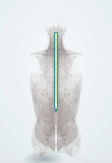
Spinal cord !
The spinal cord is the main nerve cable joining the brain to the body; it runs inside the spinal canal from the skull to about L1-L2.
Opens in the Durgical app, no sign-up needed.
What it is
The main nerve cable joining the brain to the body, running inside the spinal canal from the skull to about the L1-L2 level.
What it does
Carries movement signals from the brain and sensation back to it, and handles reflexes on its own.
Key facts
- Where
- Vertebral canal from foramen magnum (continuous with medulla) to about L1–L2 in adults; about 42–45 cm long; cervical (C4–T1) and lumbosacral (L1–S3) enlargements
- Attaches
- Denticulate ligaments (pia to dura) on each side; filum terminale to coccyx
- Function
- Ascending sensory and descending motor tracts; 31 pairs of spinal nerves; spinal reflexes; lateral horn: sympathetic preganglionic neurons T1–L2 (ciliospinal centre C8–T2), sacral parasympathetic S2–S4
Relations
- Course
- From the foramen magnum, where it continues from the medulla oblongata, down the vertebral canal to the conus medullaris at about L1–L2; the lower its roots arise, the longer and steeper their path down to their foramina, ending as the cauda equina
- In front
- Across the epidural space, the posterior longitudinal ligament over the vertebral bodies and discs; the anterior spinal artery runs in the anterior median fissure
- Behind
- Epidural fat and veins, then the laminae and ligamenta flava
- Lateral
- Denticulate ligaments to the dura, between the anterior and posterior roots, and the roots heading to the intervertebral foramina
- Superficial
- From inside out: pia mater, the subarachnoid space with CSF, arachnoid and dura mater, then the epidural space with the internal vertebral venous plexus
- Contents
- White matter in three funiculi around the grey H: posterior (fasciculus gracilis medially for the leg, cuneatus laterally for the arm, above T6), lateral (lateral corticospinal, dorsal and ventral spinocerebellar, rubrospinal; spinothalamic fibres anterolaterally) and anterior (anterior corticospinal, vestibulo-, tecto- and reticulospinal)
How to find it
It cannot be felt. Its levels are estimated from surface landmarks: vertebra prominens (C7), inferior angle of the scapula (about T7) and the line across the iliac crests (about L4, Tuffier line), well below the end of the cord. Segment under a spinous process: add about 1 in the neck, 2 at T1–T6, 3 at T7–T9; T10–T12 spines overlie the lumbar segments, L1 the sacral ones.
Examination
- ISNCSCI (ASIA) examination: 10 key muscles and 28 key sensory points per side plus an anal exam; the neurological level is the lowest segment with normal strength and sensation on both sides
- Babinski sign: stroking the outer sole makes the big toe extend and the other toes fan; an upper motor neuron sign (normal in infants)
- Hoffmann sign: flicking the nail of the middle finger makes the thumb and index flex; it suggests an upper motor neuron lesion, often cervical myelopathy
- Lhermitte sign: bending the neck forward sends an electric feeling down the spine or limbs; it points to a cervical dorsal column lesion (multiple sclerosis, myelopathy)
- Romberg test: steady with eyes open, swaying or falling with eyes closed; loss of position sense, as in dorsal column disease
- Reflexes map the segment: biceps C5–C6, brachioradialis C6, triceps C7, patellar L3–L4, Achilles S1; superficial abdominal T8–T12, cremasteric L1–L2, anal wink and bulbocavernosus S2–S4; a lost reflex marks the level, brisk reflexes lie below it
On imaging
MRI is the study of choice: T2 shows compression by disc, bone or ligament and bright signal inside the cord (oedema, myelomalacia, syrinx). CT shows fractures and the bony canal better; CT myelography is used when MRI is not possible.
Common problems
- Cervical myelopathy: slow squeezing by discs, spurs or a thick ligament, causing clumsy hands, numbness and unsteady walking.
- Spinal cord injury from fractures or dislocations, which can cause paralysis below the level.
- Inflammation (myelitis), tumours or problems with its blood supply.
How it is diagnosed
Neurological exam (strength, feeling, reflexes, balance) and MRI.
Treatment
- Cord compression with symptoms: surgery to take pressure off, such as ACDF in the neck.
- Acute injury: emergency care to stabilise the spine, then rehabilitation.
Clinical notes
- Complete vs incomplete injury: in a complete injury (ASIA A) no motor or sensory function remains in S4–S5; any sacral sparing (perianal feeling, voluntary anal squeeze) makes it incomplete, with a better outlook
- Central cord syndrome, the commonest incomplete pattern: an older person with a narrowed, stiff cervical spine falls and hyperextends the neck; arms weaker than legs, hands worst, variable sensory and bladder loss
- Brown-Séquard syndrome (hemisection, e.g. a stab wound): same-side weakness and loss of vibration and position sense, opposite-side loss of pain and temperature starting one or two segments below the lesion; anterior cord syndrome (anterior spinal artery): paralysis and pain loss with dorsal columns spared
- Cervical spondylotic myelopathy, the commonest cord disorder in older adults: the clumsy hand, broad-based gait, Hoffmann sign and brisk reflexes build up slowly, often with little neck pain
- Conus vs cauda equina: a conus lesion starts suddenly and symmetrically, with early sphincter failure, little pain and mixed upper and lower motor neuron signs; cauda equina compression is often asymmetric, painful and purely lower motor neuron
Development
In the third fetal month the cord fills the whole canal; the column then outgrows it, so the conus lies at about L3 at birth and reaches the adult level in infancy. A tight filum that holds it low (tethered cord) stretches the cord as the child grows.
Related conditions
- Morquio syndrome (mucopolysaccharidosis type IV)
- Multiple sclerosis
- Spinal cord injury
- Degenerative cervical myelopathy
- Spinal epidural abscess
- Spina bifida (myelomeningocele)
- Neurogenic shock
- Decompression sickness and diving injuries
- Malignant spinal cord compression
- Tetanus
- Poliomyelitis and acute flaccid paralysis
- Amyotrophic lateral sclerosis
- Subacute combined degeneration (vitamin B12)
- Autonomic dysreflexia
- Spinal muscular atrophy
- Transverse myelitis
- Neuromyelitis optica spectrum disorder
- Syringomyelia and Chiari malformation
- Friedreich ataxia
- HTLV-1-associated myelopathy / tropical spastic paraparesis
- Major trauma (polytrauma): ATLS primary survey
- Cervical spine fracture
- Cervical Spondylosis
Test yourself
Think of the answer, then tap the question to see it.
At what vertebral level does the adult spinal cord end?
At about L1–L2, as the conus medullaris; it is continuous with the medulla at the foramen magnum.
Which artery feeds the lower spinal cord, and where does it usually arise?
The artery of Adamkiewicz, usually on the left between T9 and T12; the anterior spinal artery supplies about the front two-thirds of the cord.
What is the pattern of Brown-Séquard syndrome?
Cord hemisection: same-side weakness and loss of vibration and position sense; opposite-side loss of pain and temperature.
Which incomplete spinal cord injury is most common, and what is its typical picture?
Central cord syndrome: an older person with a stiff, narrow cervical spine hyperextends the neck; arms weaker than legs, hands worst.
What makes a spinal cord injury incomplete on the ASIA scale?
Any sacral sparing (perianal sensation or voluntary anal squeeze in S4–S5); a complete injury (ASIA A) has none, and incomplete ones have a better outlook.
Educational information, not medical advice. For any symptom, see a health professional.
Its names
- Official Latin (TA2)
- medulla spinalis; substantia alba medullae spinalis TA2 6049
- Official Spanish (DeCS)
- Médula espinal DeCS D013116
- English (TA2)
- Spinal cord
- Also called
- white columns of spinal cord
Related
- Dura (dural sac)
- Conus medullaris
- Grey matter of the cord
- Vertebral artery
- Posterior intercostal arteries
- Anterior spinal artery
- Posterior spinal arteries
- Artery of Adamkiewicz
Sources: Terminologia Anatomica (TA2); DeCS; Neuroanatomy, Spinal Cord; Neuroanatomy, Spinal Cord Morphology; Tratado de anatomía humana (9.ª ed.), tomo II, Sistema nervioso central: cerebro, núcleos grises centrales, vías de conducción y circulación cerebral; Tratado de anatomía humana (9.ª ed.), tomo II, Libro V Sistema nervioso central, sección 1: Médula espinal.
Spotted a mistake?We are always improving for the community. Tell us what is wrong and our team will review it.Report a mistake
Representative model, for educational purposes only.