Brachioradialis !
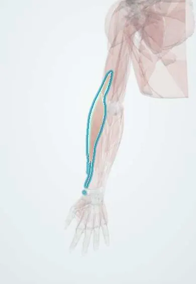
The brachioradialis runs along the thumb side of the forearm from above the elbow to the radius; it bends the elbow, strongest with the thumb up.
Opens in the Durgical app, no sign-up needed.
What it is
The forearm muscle on the thumb side, from above the elbow to the end of the radius.
What it does
Bends the elbow, strongest with the thumb pointing up.
Key facts
- Origin
- Upper lateral supracondylar ridge of the humerus
- Insertion
- Base of the radial styloid process
- Action
- Flexes the elbow, strongest in mid-pronation
- Nerve
- Radial nerve (C5–C6)
- Blood supply
- Radial recurrent artery
Relations
- Deep
- Supinator and extensor carpi radialis longus (ECRL); the superficial radial nerve runs under it down the forearm and emerges between its tendon and ECRL
- Medial
- Pronator teres across the cubital fossa; lower down the radial artery runs along its medial edge, between it and flexor carpi radialis
- Behind
- Extensor carpi radialis longus (ECRL) and brevis (ECRB), forming with it the mobile wad of the lateral forearm
- Superficial
- Skin with the cephalic vein and the lateral cutaneous nerve of the forearm
How to find it
With the forearm in mid-position (thumb up) and the elbow flexed against resistance, it stands out as the bulge along the thumb side of the upper forearm.
Examination
- Brachioradialis reflex: the tendon is tapped just above the radial styloid; reduced response suggests a C6 root or radial nerve lesion, a brisk one an upper motor neuron lesion
- Resisted elbow flexion in mid-pronation: weakness with a strong biceps suggests a radial nerve lesion at or above the spiral groove
On imaging
On ultrasound and MRI it is the most superficial muscle of the lateral forearm and a landmark: the radial nerve lies between it and brachialis at the elbow, and the superficial radial nerve runs under it. In distal radius fractures, its pull on the radial styloid helps shorten the radius. MRI shows strain or, after radial nerve injury, denervation.
Clinical notes
- Wartenberg syndrome (cheiralgia paresthetica): the superficial radial nerve is pinched where it emerges between the brachioradialis and ECRL tendons, often by tight wristbands
- Deforming force in distal radius fractures, pulling the distal fragment up; its insertion is sometimes released to help reduction
- Transfer of brachioradialis to FPL restores key pinch in tetraplegia when elbow flexion is preserved
Development
A dorsal (extensor) mass muscle that ends up working as an elbow flexor; this origin is why the radial nerve supplies it, unlike the other elbow flexors.
Test yourself
Think of the answer, then tap the question to see it.
In which forearm position is the brachioradialis strongest, and what does it do?
Mid-pronation (thumb up); it flexes the elbow.
Why does the brachioradialis still work in posterior interosseous nerve palsy?
It is supplied by the radial nerve above its division into superficial and deep branches.
What is an inverted supinator reflex, and what does it indicate?
Tapping the brachioradialis tendon gives finger flexion without elbow flexion; it points to a C5–C6 cord lesion.
What is Wartenberg syndrome (cheiralgia paresthetica)?
Compression of the superficial radial nerve where it emerges between the brachioradialis and ECRL tendons, often by tight wristbands.
Educational information, not medical advice. For any symptom, see a health professional.
Its names
- Official Latin (TA2)
- musculus brachioradialis TA2 2496
- Official Spanish (DeCS)
- Músculo braquiorradial
- English (TA2)
- Brachioradialis
Related
Sources: Terminologia Anatomica (TA2); Anatomy, Shoulder and Upper Limb, Forearm Brachioradialis Muscle.
Spotted a mistake?We are always improving for the community. Tell us what is wrong and our team will review it.Report a mistake
Representative model, for educational purposes only.