Radial nerve !
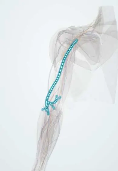
The radial nerve winds around the back of the humerus; it moves the muscles that straighten the elbow, wrist and fingers.
Opens in the Durgical app, no sign-up needed.
What it is
The nerve that spirals around the back of the humerus to the outer elbow.
What it does
Powers the muscles that straighten the elbow, wrist and fingers.
Key facts
- Roots
- C5–T1, posterior cord (some texts: C5–C8)
- Moves
- Triceps, anconeus, brachioradialis, ECRL; via the deep branch/PIN: ECRB, supinator and the forearm extensors
- Feeling from
- Posterior arm and forearm; dorsum of the lateral hand and lateral 3½ digits (proximal parts)
Relations
- Course
- Behind the third part of the axillary artery, through the triangular interval with the deep brachial artery, along the spiral groove between the lateral and medial heads of triceps, through the lateral intermuscular septum, then between brachialis and brachioradialis to divide in front of the lateral epicondyle
- Behind
- The lateral head of triceps, which covers it in the spiral groove
- Deep
- The humeral shaft in the spiral groove, against which it is pressed in fractures and in sleep palsy
- Medial
- Brachialis, above and in front of the elbow
- Lateral
- Brachioradialis and extensor carpi radialis longus, which it supplies before dividing
How to find it
It cannot be felt in the arm. In a posterior approach to the humerus it is found crossing the shaft in the spiral groove between the lateral and medial heads of triceps, with the deep brachial vessels; in a lateral approach it is found above the elbow between brachialis and brachioradialis and traced upward.
Examination
- Wrist drop: the wrist and the fingers at the MCP joints cannot be lifted; finger IP extension is kept (interossei and lumbricals), so MCP extension is tested with the wrist supported
- Brachioradialis reflex (C6) lost with a normal triceps reflex (C7) fits a spiral groove lesion; both lost suggests a lesion in the axilla
- Pinprick on the dorsum of the first web space, the area supplied by the radial nerve alone
On imaging
It lies in the radial groove of the humerus, so a shaft fracture on radiographs, classically a distal-third spiral fracture (Holstein-Lewis), raises concern for it. Ultrasound follows the nerve along the humerus to show if it is intact, swollen, or caught in the fracture or callus, and shows its split into superficial and deep branches at the elbow. MRI shows denervation.
Common problems
- Injured with fractures of the humerus shaft, where it wraps around the bone: wrist drop.
- 'Saturday night palsy' from pressure on the arm during deep sleep.
- Radial tunnel syndrome: aching in the forearm from compression near the supinator.
How it is diagnosed
Exam of wrist and finger lifting strength and of feeling on the back of the hand. EMG and nerve conduction studies.
Treatment
- A wrist splint and watching for recovery; most pressure and fracture-related injuries recover.
- Nerve exploration, repair or tendon transfers when it does not recover.
Clinical notes
- Saturday night palsy vs crutch palsy: an arm hung over a chair back compresses the nerve in the spiral groove (triceps spared); crutch pressure in the axilla compresses it above the triceps branches (triceps weak)
- Humeral shaft fractures are the commonest cause of radial nerve palsy; most closed-fracture palsies are neurapraxias that recover, while a palsy appearing after manipulation raises concern for entrapment
- At risk in posterior and lateral approaches to the humerus, plating of the shaft and lateral external fixator pins
- Radial nerve vs C7 radiculopathy vs PIN palsy: C7 also weakens triceps and wrist flexion; PIN palsy keeps wrist extension (in radial deviation) and sensation
Anatomical variants
- Its deep branch may pass between or through the two heads of supinator, under an arcade of Frohse of variable thickness
Development
It carries the posterior cord into the dorsal (extensor) muscle mass. The limb's outward rotation in week 7 turns that surface backward, so all its muscles belong to the posterior compartments, brachioradialis included, even though that muscle bends the elbow.
Memory aid
Radial nerve muscles, 'BEST': Brachioradialis, Extensors, Supinator, Triceps.
Related conditions
Test yourself
Think of the answer, then tap the question to see it.
Through which space does the radial nerve leave the axilla, and with which vessel?
The triangular interval (below teres major, between the long head of triceps and the humerus), with the deep brachial artery.
What is a Holstein-Lewis fracture, and why does it matter for the radial nerve?
A spiral fracture of the distal third of the humeral shaft; it can trap the radial nerve where it pierces the lateral intermuscular septum.
Why is the triceps usually spared in a radial nerve lesion at the spiral groove?
Its branches leave higher, in the axilla and upper arm; only an axillary lesion (such as crutch pressure) weakens the triceps too.
What is the classic sign of a radial nerve palsy, and why is finger IP extension kept?
Wrist drop: the wrist and MCP joints cannot be extended; IP extension is kept by the interossei and lumbricals.
Where is the area supplied by the radial nerve alone tested?
By pinprick on the dorsum of the first web space.
Educational information, not medical advice. For any symptom, see a health professional.
Its names
- Official Latin (TA2)
- nervus radialis TA2 6431
- Official Spanish (DeCS)
- Nervio radial
- English (TA2)
- Radial nerve
Related
- Deep brachial artery
- Cubital fossa
- Triceps (long head)
- Triceps (lateral head)
- Triceps (medial head)
- Anconeus
- Brachioradialis
- ECRL
Sources: Terminologia Anatomica (TA2); Anatomy, Shoulder and Upper Limb, Radial Nerve; 8.5 Development of the Appendicular Skeleton (OpenStax (Rice University), 2022) · adapted, CC BY 4.0; Anatomy of the Human Body (20th ed.): The Branchial Region (Lea & Febiger (Gray's Anatomy, 1918), 1918) · adapted, Public domain.
Spotted a mistake?We are always improving for the community. Tell us what is wrong and our team will review it.Report a mistake
Representative model, for educational purposes only.