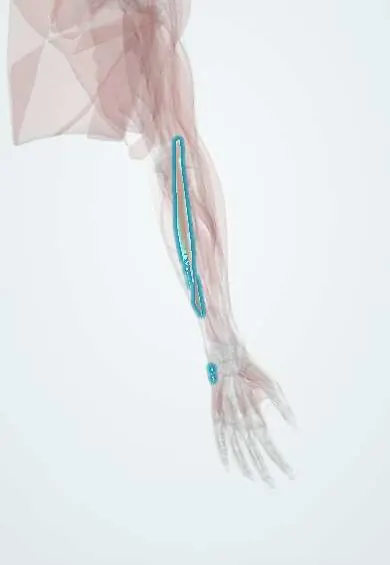
ECU !
The extensor carpi ulnaris runs along the little-finger side of the back of the forearm; it lifts the wrist and tilts it toward the little finger.
Official name: Extensor carpi ulnaris
Opens in the Durgical app, no sign-up needed.
What it is
A wrist extensor on the little-finger side, to the base of the 5th metacarpal.
What it does
Lifts the wrist and tilts it toward the little finger; its sheath helps steady the TFCC.
Key facts
- Origin
- Lateral epicondyle and posterior border of the ulna
- Insertion
- Base of the 5th metacarpal
- Action
- Extends and ulnarly deviates the wrist
- Nerve
- Posterior interosseous nerve (C7–C8)
- Blood supply
- Ulnar artery, posterior interosseous artery
Relations
- Lateral
- Extensor digiti minimi
- Medial
- The subcutaneous posterior border of the ulna, with flexor carpi ulnaris on its other side
- Deep
- The ulna; at the wrist, the ulnar styloid groove and the triangular fibrocartilage complex (TFCC)
- Course
- Through the sixth compartment in its subsheath, then across the ulnar side of the carpus to the base of the 5th metacarpal
How to find it
With the wrist extended and tilted toward the little finger, the tendon is felt in the groove next to the ulnar styloid and just before the base of the 5th metacarpal; feeling it while the forearm turns from pronation to supination can reveal instability.
Examination
- ECU synergy test: with the forearm supinated, resisted radial abduction of the thumb makes ECU contract; pain along the dorsal ulnar wrist points to ECU tendinitis rather than a TFCC tear
- Subluxation test: active supination with wrist flexion and ulnar deviation; a palpable snap of the tendon indicates a subsheath injury
On imaging
Ultrasound and MRI show the tendon in the ulnar head groove, held by its own subsheath in the sixth compartment. Classic findings: tenosynovitis (sheath fluid) and instability, with dynamic ultrasound in supination and ulnar deviation showing it slip out of the groove. MRI also assesses the triangular fibrocartilage complex. Radiographs show only bony causes.
Clinical notes
- ECU tendinitis and instability are common mimics of TFCC tears in ulnar-sided wrist pain
- Wrist arthroscopy portals 6R and 6U are named for their position on either side of the ECU tendon; the dorsal branch of the ulnar nerve is at risk at 6U
- In rheumatoid arthritis the tendon can slip forward and lose its extensor action, adding to radial deviation of the wrist
Anatomical variants
- Accessory slips running on to the little finger's extensor mechanism or the 5th metacarpal are described
Test yourself
Think of the answer, then tap the question to see it.
What holds the ECU (extensor carpi ulnaris) tendon in place at the wrist?
Its own fibro-osseous subsheath in the groove beside the ulnar styloid, under the extensor retinaculum (sixth compartment).
What does snapping of the ECU tendon on supination, wrist flexion and ulnar deviation mean?
A subsheath tear, seen in tennis and golf.
How does the ECU synergy test help in ulnar-sided wrist pain?
Forearm supinated, resisted radial abduction of the thumb makes the ECU contract; dorsal ulnar pain points to ECU tendinitis rather than a TFCC tear.
Educational information, not medical advice. For any symptom, see a health professional.
Its names
- Official Latin (TA2)
- extensor ulnaris carpi TA2 2507
- Official Spanish (DeCS)
- Músculo extensor ulnar del carpo
- English (TA2)
- Extensor carpi ulnaris
Related
- Posterior interosseous nerve
- Interosseous arteries
- Humerus
- 5th metacarpal
- Triquetrum
- Ulna
- Common extensor tendon
- Wrist extensor tendons
Sources: Terminologia Anatomica (TA2); Anatomy, Shoulder and Upper Limb, Forearm Extensor Carpi Ulnaris Muscle; Anatomy, Shoulder and Upper Limb, Wrist Extensor Muscles.
Spotted a mistake?We are always improving for the community. Tell us what is wrong and our team will review it.Report a mistake
Representative model, for educational purposes only.