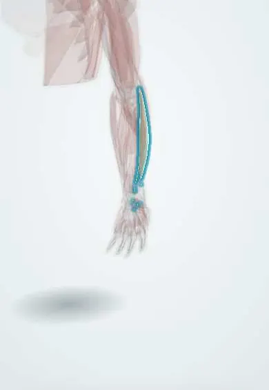
Extensor digitorum !
The extensor digitorum runs down the back of the forearm with tendons to four fingers; it straightens the fingers and helps lift the wrist.
Opens in the Durgical app, no sign-up needed.
What it is
The main finger-straightening muscle on the back of the forearm.
What it does
Straightens the fingers and helps lift the wrist.
Key facts
- Origin
- Lateral epicondyle
- Insertion
- Extensor expansions of digits 2–5
- Action
- Extends the fingers (mainly at the MCP joints) and the wrist
- Nerve
- Posterior interosseous nerve (C7–C8)
- Blood supply
- Posterior interosseous artery
Relations
- Lateral
- Extensor carpi radialis brevis (ECRB)
- Medial
- Extensor digiti minimi, then the extensor carpi ulnaris (ECU)
- Deep
- The deep extensor layer: supinator, APL, EPB, EPL and extensor indicis, with the posterior interosseous nerve and artery between the layers
- Course
- Through the fourth compartment and across the back of the hand; over each finger it forms the extensor mechanism, with a central slip to the middle phalanx and lateral bands to the distal phalanx
How to find it
With the fingers straightened at the knuckles against resistance, its tendons fan out visibly on the back of the hand; the belly is felt in the middle of the back of the forearm below the lateral epicondyle.
Examination
- Resisted MCP extension: loss of finger extension with preserved, radially deviated wrist extension (finger drop without wrist drop) indicates posterior interosseous nerve palsy
- Elson test: with the PIP joint bent over a table edge, resisted PIP extension that makes the fingertip rigid indicates a central slip rupture
On imaging
Ultrasound shows its tendons in the fourth extensor compartment and over the metacarpal heads, centred by the sagittal bands; making a fist shows a tendon slipping off the knuckle after a sagittal band tear (boxer's knuckle). In mallet finger a lateral finger radiograph looks for a fragment from the distal phalanx base; ultrasound or MRI for tears.
Clinical notes
- Central slip injury leads to a boutonnière deformity; loss of the terminal tendon to a mallet finger
- Sagittal band rupture lets the tendon slip off the knuckle toward the ulnar side (seen after punching); in rheumatoid arthritis it adds to ulnar drift
- In permanent radial nerve palsy, finger extension is restored by transferring FCR or FCU to its tendons
Anatomical variants
- Tendon slips are often doubled or missing; the little-finger tendon may be absent, replaced by a junctura, leaving extension to extensor digiti minimi
- Extensor digitorum brevis manus, a small accessory muscle on the back of the hand, can be mistaken for a ganglion
Development
Comes from the dorsal muscle mass of the limb bud. When the upper limb turns outward by about 90° in week 7, this mass ends up on the back of the forearm and the thumb on the lateral side, so the extensors lie posterior and take the radial nerve.
Related conditions
Test yourself
Think of the answer, then tap the question to see it.
Which joints does the extensor digitorum mainly extend, and what else is needed for IP extension?
Mainly the MCP joints; IP extension also needs the lumbricals and interossei through the extensor mechanism.
What is the pattern of posterior interosseous nerve palsy at the extensor digitorum?
Finger drop without wrist drop: loss of MCP extension with preserved, radially deviated wrist extension.
Which deformity follows a central slip injury of the extensor digitorum mechanism?
A boutonnière deformity; loss of the terminal tendon gives a mallet finger.
Educational information, not medical advice. For any symptom, see a health professional.
Its names
- Official Latin (TA2)
- extensor digitorum TA2 2500
- Official Spanish (DeCS)
- Músculo extensor de los dedos
- English (TA2)
- Extensor digitorum
Related
Sources: Terminologia Anatomica (TA2); Anatomy, Shoulder and Upper Limb, Hand Compartments; Anatomy, Shoulder and Upper Limb, Wrist Extensor Muscles; 8.5 Development of the Appendicular Skeleton (OpenStax (Rice University), 2022) · adapted, CC BY 4.0.
Spotted a mistake?We are always improving for the community. Tell us what is wrong and our team will review it.Report a mistake
Representative model, for educational purposes only.