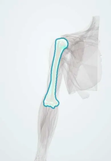
Humerus !
The humerus is the upper arm bone, from shoulder to elbow; it forms the ball of the shoulder and carries the main arm muscles.
Opens in the Durgical app, no sign-up needed.
What it is
The long bone of the upper arm, from the ball of the shoulder to the elbow.
What it does
Carries the arm muscles and forms the ball of the shoulder and the top of the elbow.
Key facts
- Joints with
- Scapula (glenoid), radius (capitulum), ulna (trochlea)
- Landmarks
- Head, anatomical and surgical necks, greater and lesser tubercles, intertubercular sulcus, deltoid tuberosity, radial groove, medial and lateral epicondyles, capitulum, trochlea; coronoid and radial fossae (front), olecranon fossa (back); medial and lateral supracondylar ridges
Relations
- Above
- Head against the glenoid; supraspinatus to the top of the greater tubercle, infraspinatus and teres minor behind it, subscapularis to the lesser tubercle
- In front
- Long biceps tendon in the intertubercular groove under the transverse humeral ligament; distally the brachialis covers the front of the shaft
- Behind
- Radial nerve and deep brachial artery spiral down in the radial groove between the origins of the lateral and medial heads of the triceps; the axillary nerve and posterior circumflex humeral artery wind around the surgical neck
- Medial
- Coracobrachialis at mid-shaft; the brachial artery and median nerve run along the medial side; the ulnar nerve lies in its groove behind the medial epicondyle
- Lateral
- Deltoid tuberosity at mid-shaft; brachioradialis and ECRL from the lateral supracondylar ridge; the common extensor tendon from the lateral epicondyle
How to find it
The greater tubercle is the most lateral bony point of the shoulder, felt below the acromion and rolling under the fingers as the arm is rotated; the intertubercular groove lies in front of it. At the elbow both epicondyles are subcutaneous: with the elbow extended they line up with the tip of the olecranon, and with it flexed to 90° the three points form a triangle.
On imaging
Shoulder: true AP, scapular Y and axillary views; proximal fractures are grouped by the number of displaced parts (Neer: head, greater tubercle, lesser tubercle, shaft). Child's elbow: on the lateral view the anterior humeral line should cross the middle third of the capitulum, and a visible posterior fat pad after trauma suggests an occult fracture.
Common problems
- Fracture near the shoulder (proximal humerus), common in older adults after a fall.
- Shaft fracture, which can stretch the radial nerve (wrist drop).
- Fractures above the elbow (supracondylar), most often in children.
How it is diagnosed
X-rays in two planes; CT for complex fractures near the joints. Nerve and pulse checks for every fracture.
Treatment
- Sling, brace or cast for fractures that are stable and well aligned.
- Surgery with plates, rods or pins for displaced fractures.
- Shoulder replacement when the humeral head is broken into many pieces or worn out.
Clinical notes
- Supracondylar fractures can cause forearm compartment syndrome and, if missed, Volkmann ischaemic contracture; malunion leaves a cubitus varus (gunstock deformity)
- Anterior shoulder dislocation dents the posterolateral head against the glenoid rim (Hill–Sachs lesion); posterior dislocation leaves a reverse Hill–Sachs lesion on the front of the head
- In elbow dislocation in children the medial epicondyle can be avulsed and trapped inside the joint
- The epicondyle–olecranon triangle stays normal in a supracondylar fracture but is distorted in an elbow dislocation
Anatomical variants
- Supracondylar process: a small bony spur above the medial epicondyle in about 1% of people; a band from it (ligament of Struthers) can compress the median nerve and brachial artery
- Septal aperture: a hole between the olecranon and coronoid fossae, which can look like a lytic lesion on X-ray
Development
Elbow ossification centres appear in a fixed order (CRITOE), roughly every two years from about age 1: capitulum, radial head, internal (medial) epicondyle, trochlea, olecranon, external (lateral) epicondyle; earlier in girls. A trochlear centre with no medial epicondyle in its usual place suggests the epicondyle is trapped in the joint. The proximal physis gives most of its growth in length.
Memory aid
CRITOE for the elbow centres: Capitulum, Radial head, Internal epicondyle, Trochlea, Olecranon, External epicondyle.
Related conditions
- Osteosarcoma
- Shoulder dislocation
- Lateral epicondylitis (tennis elbow)
- Supracondylar humerus fracture
- Proximal humerus fracture
- Humeral shaft fracture
- Medial epicondylitis (golfer's elbow)
- Elbow dislocation
Test yourself
Think of the answer, then tap the question to see it.
Which three nerves lie directly on the humerus, and where?
The axillary nerve at the surgical neck, the radial nerve in the radial groove of the shaft and the ulnar nerve behind the medial epicondyle.
What is a Holstein–Lewis fracture of the humerus, and which deficit does it cause?
A spiral fracture of the distal third of the humeral shaft with radial nerve palsy, causing wrist drop.
Which structures are endangered by an extension-type supracondylar fracture of the humerus in a child?
The brachial artery and the median nerve, especially its anterior interosseous branch.
Which muscles attach to the lips and floor of the intertubercular groove of the humerus?
Pectoralis major on the lateral lip, teres major on the medial lip and latissimus dorsi in the floor.
What does the CRITOE mnemonic give for the elbow?
The order in which the elbow ossification centres appear: capitulum, radial head, internal (medial) epicondyle, trochlea, olecranon, external (lateral) epicondyle.
Educational information, not medical advice. For any symptom, see a health professional.
Its names
- Official Latin (TA2)
- humerus TA2 1180
- Official Spanish (DeCS)
- Húmero
- English (TA2)
- Humerus
- Also called
- upper arm bone
Related
- Circumflex humeral arteries
- Deep brachial artery
- Anconeus
- Biceps (long head)
- Long biceps tendon
- Biceps (short head)
- Biceps tendon sheath
- Brachialis
Sources: Terminologia Anatomica (TA2); Anatomy, Shoulder and Upper Limb, Humerus.
Spotted a mistake?We are always improving for the community. Tell us what is wrong and our team will review it.Report a mistake
Representative model, for educational purposes only.