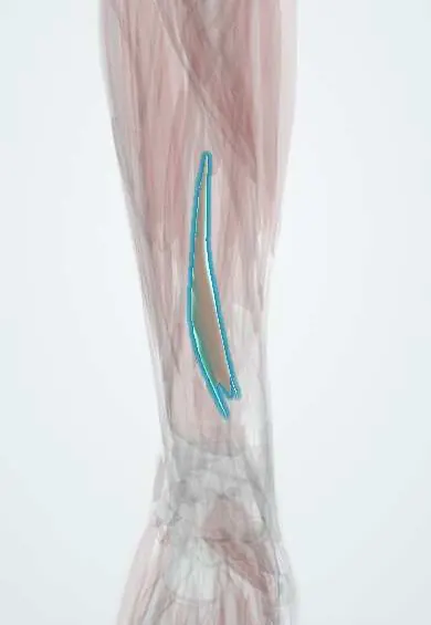
EPL !
The extensor pollicis longus is a deep forearm muscle whose tendon reaches the thumb tip; it lifts the thumb up and back.
Official name: Extensor pollicis longus
Opens in the Durgical app, no sign-up needed.
What it is
A deep forearm muscle whose tendon goes to the tip of the thumb.
What it does
Lifts the thumb up and back.
Key facts
- Origin
- Middle third of the posterior ulna and interosseous membrane
- Insertion
- Base of the distal phalanx of the thumb
- Action
- Extends the whole thumb (IP joint); hooks round Lister's tubercle
- Nerve
- Posterior interosseous nerve (C7–C8)
- Blood supply
- Posterior interosseous artery
Relations
- Course
- From the back of the ulna through the third compartment on the ulnar side of Lister's tubercle; it then crosses the ECRL and ECRB tendons obliquely to reach the thumb
- Medial
- Extensor indicis in the deep layer of the forearm; the extensor digitorum tendons in the fourth compartment
- Lateral
- APL and EPB in the deep extensor layer
- Deep
- The dorsal radius at Lister's tubercle; distally the ECRL and ECRB tendons where it crosses them
How to find it
With the palm flat on a table, lifting the thumb straight up off it tightens the tendon, which is seen as the dorsal edge of the anatomical snuffbox and can be traced back to Lister's tubercle.
Examination
- Retropulsion test: inability to lift the thumb off the table with the hand flat indicates EPL rupture, a cut or posterior interosseous nerve palsy
On imaging
Ultrasound follows it around Lister's tubercle in the third compartment; a rupture is a gap with a retracted end. Classic: late rupture weeks after an undisplaced distal radius fracture, or wear from a protruding dorsal screw. Radiographs show the fracture or screw; ultrasound or MRI shows the tendon. It forms the ulnar border of the anatomical snuffbox.
Clinical notes
- Extensor indicis to EPL transfer is the classic reconstruction after rupture
- Drill bits or screws that protrude from the back of the radius during volar plating can wear through the tendon
- Attrition rupture also occurs in rheumatoid arthritis
Anatomical variants
- Rare tendon anomalies, such as a doubled tendon or an extra slip to the index finger, have been described and classified
Related conditions
Test yourself
Think of the answer, then tap the question to see it.
Why does the EPL (extensor pollicis longus) rupture weeks after a distal radius fracture?
From wear and poor blood supply where it turns round Lister's tubercle, often after an undisplaced fracture.
Which test reliably detects EPL rupture, and why is IP extension not enough?
Retropulsion: lifting the thumb off a flat table; the thenar muscles can still extend the IP joint weakly.
What is the classic reconstruction after EPL rupture?
Transfer of the extensor indicis to the EPL.
Educational information, not medical advice. For any symptom, see a health professional.
Its names
- Official Latin (TA2)
- extensor longus pollicis TA2 2516
- Official Spanish (DeCS)
- Músculo extensor largo del pulgar
- English (TA2)
- Extensor pollicis longus
Related
Sources: Terminologia Anatomica (TA2); Anatomy, Shoulder and Upper Limb, Hand Extensor Pollicis Longus Muscle; Anatomy, Shoulder and Upper Limb: Hand Anatomical Snuff Box.
Spotted a mistake?We are always improving for the community. Tell us what is wrong and our team will review it.Report a mistake
Representative model, for educational purposes only.