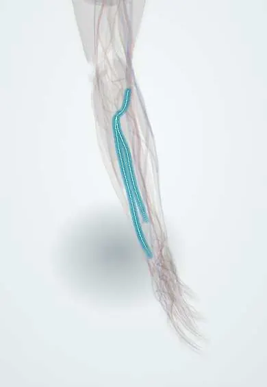
Posterior interosseous nerve !
The posterior interosseous nerve is the deep branch of the radial nerve in the forearm; it moves the muscles that straighten the fingers and thumb.
Opens in the Durgical app, no sign-up needed.
What it is
The deep branch of the radial nerve that passes through the supinator; it can be squeezed there (radial tunnel) and must be protected in distal biceps and radial head surgery.
What it does
Powers the finger and thumb extensors.
Key facts
- Roots
- C7–C8, deep branch of the radial nerve
- Moves
- Supinator, ECRB, extensor digitorum, EDM, ECU, APL, EPB, EPL, extensor indicis
- Feeling from
- No skin (wrist joint only)
Relations
- Course
- Arises from the radial nerve in front of the lateral epicondyle, passes under the edge of ECRB and the arcade of Frohse into supinator, winds round the lateral side of the radial neck between its layers, emerges in the back of the forearm, runs on the interosseous membrane and ends in the floor of the fourth extensor compartment
- In front
- At the radial tunnel, the radial recurrent vessels (leash of Henry) and the edge of extensor carpi radialis brevis
- Deep
- The neck of the radius, with only the deep layer of supinator between them
- Superficial
- The superficial extensors (extensor digitorum, extensor digiti minimi, ECU) once it leaves supinator
How to find it
Not palpable. In a posterior (Thompson) approach it is found as it leaves the distal edge of supinator between ECRB and extensor digitorum; at the wrist its terminal branch lies on the floor of the fourth extensor compartment, under the extensor digitorum tendons.
Examination
- Resisted finger extension at the MCP joints with the wrist supported: weakness with intact sensation and wrist extension points to the PIN
- Resisted middle finger extension: pain over the radial tunnel, distal to the lateral epicondyle, suggests radial tunnel syndrome rather than lateral epicondylitis
- Tenderness over the supinator, a few finger-breadths distal to the lateral epicondyle, rather than at the epicondyle itself
On imaging
Ultrasound follows it from the radial nerve split into the supinator under the arcade of Frohse: look for swelling just before the arcade or a compressing mass (ganglion, lipoma, elbow synovitis). MRI shows denervation oedema, later fatty atrophy, of the extensors. After a Monteggia injury or radial head dislocation, radiographs show the injury that can stretch it.
Common problems
- Injured with fractures of the humerus shaft, where it wraps around the bone: wrist drop.
- 'Saturday night palsy' from pressure on the arm during deep sleep.
- Radial tunnel syndrome: aching in the forearm from compression near the supinator.
How it is diagnosed
Exam of wrist and finger lifting strength and of feeling on the back of the hand. EMG and nerve conduction studies.
Treatment
- A wrist splint and watching for recovery; most pressure and fracture-related injuries recover.
- Nerve exploration, repair or tendon transfers when it does not recover.
Clinical notes
- Same tunnel, two pictures: radial tunnel syndrome is pain without weakness; PIN syndrome is weakness with little pain
- At risk with radial head and neck fractures, two-incision distal biceps repair, plating of the proximal radius and posterior forearm approaches
- Masses at the elbow (lipoma, ganglion, rheumatoid synovitis) can compress it and present as painless finger drop
- Its terminal branch in the fourth compartment is divided in wrist denervation and can serve as a graft for digital nerves
Anatomical variants
- The arcade of Frohse, the upper edge of supinator, is often tendinous in adults, and the nerve may pass between or through the two heads of supinator
Memory aid
Radial tunnel compression sites, 'FREAS': Fibrous bands, Recurrent radial vessels (leash of Henry), ECRB edge, Arcade of Frohse, Supinator distal edge.
Related conditions
Test yourself
Think of the answer, then tap the question to see it.
How does posterior interosseous nerve palsy look?
Fingers and thumb drop at the MCP joints, but the wrist still extends in radial deviation (ECRL spared, ECU lost) and sensation is normal.
Which fracture-dislocation classically injures the posterior interosseous nerve?
The Monteggia fracture-dislocation: the dislocated radial head stretches the nerve.
Under which structure does the posterior interosseous nerve enter supinator?
The arcade of Frohse, the upper edge of supinator, often tendinous in adults.
How does radial tunnel syndrome differ from posterior interosseous nerve syndrome?
Same tunnel, two pictures: radial tunnel syndrome is pain without weakness; PIN syndrome is weakness with little pain.
Why is the forearm pronated in lateral approaches to the radial head and neck?
Pronation moves the posterior interosseous nerve away from the surgical field.
Educational information, not medical advice. For any symptom, see a health professional.
Its names
- Official Latin (TA2)
- ramus profundus nervi radialis TA2 6436
- Official Spanish (DeCS)
- Ramo profundo del nervio radial; nervio interóseo posterior del antebrazo
- English (TA2)
- Posterior interosseous nerve
Related
Sources: Terminologia Anatomica (TA2); Anatomy, Shoulder and Upper Limb, Radial Nerve; Anatomy, Shoulder and Upper Limb, Forearm Nerves.
Spotted a mistake?We are always improving for the community. Tell us what is wrong and our team will review it.Report a mistake
Representative model, for educational purposes only.