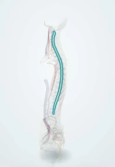
Grey matter of the cord !
The grey matter is the butterfly-shaped core of the spinal cord, made of nerve cell bodies; its front horns signal muscles, its back horns receive sensation.
Official name: Grey matter of spinal cord
Opens in the Durgical app, no sign-up needed.
What it is
The butterfly-shaped core of the spinal cord, made of nerve cell bodies (the horns), inside the white matter.
What it does
The front horns send signals to the muscles; the back horns receive sensation.
Key facts
- Where
- H-shaped central core of the cord around the central canal; anterior and posterior horns, lateral horn at T1–L2
- Function
- Anterior horn: lower motor neurons; posterior horn: sensory relay; lateral horn (T1–L2): sympathetic preganglionic neurons; S2–S4: parasympathetic
Relations
- In front
- Anterior horn, from which the ventral rootlets leave through the anterolateral sulcus; the anterior median fissure separates the two halves
- Behind
- Tip of the posterior horn with the substantia gelatinosa (lamina II), capped by the dorsolateral (Lissauer) tract where pain fibres enter
- Superficial
- White matter all around: the dorsal columns between the posterior horns, the lateral and anterior funiculi outside the horns
- Contents
- Central canal in the grey commissure; motor neuron pools in the anterior horn, with trunk muscles medial, limb muscles lateral and flexors behind extensors
How to find it
On a cross-section it is the central H- or butterfly-shaped area around the central canal, surrounded by white matter. Its shape gives the level: broad anterior horns in the cervical and lumbosacral enlargements, a small lateral horn from T1 to L2, and a large grey area with little white matter in the sacral cord.
On imaging
On axial T2 MRI the grey matter shows as a slightly brighter butterfly. Two bright dots in the anterior horns ('owl eyes' or 'snake eyes') suggest anterior horn damage, as in spinal cord infarction or compressive myelopathy.
Common problems
- Cervical myelopathy: slow squeezing by discs, spurs or a thick ligament, causing clumsy hands, numbness and unsteady walking.
- Spinal cord injury from fractures or dislocations, which can cause paralysis below the level.
- Inflammation (myelitis), tumours or problems with its blood supply.
How it is diagnosed
Neurological exam (strength, feeling, reflexes, balance) and MRI.
Treatment
- Cord compression with symptoms: surgery to take pressure off, such as ACDF in the neck.
- Acute injury: emergency care to stabilise the spine, then rehabilitation.
Clinical notes
- Syringomyelia: a fluid cavity in the centre of the cord cuts the crossing pain fibres first, giving a cape-like loss of pain and temperature over the shoulders and arms with touch spared; hand wasting follows when it reaches the anterior horns
- Lower motor neuron diseases of the anterior horn: poliomyelitis (asymmetric flaccid paralysis), spinal muscular atrophy (infants and children), ALS (with upper motor neuron signs as well)
- A lesion of the T1–T2 lateral horn interrupts the sympathetic outflow to the head, a central cause of Horner syndrome
- Loss of the sacral parasympathetic nucleus (S2–S4, in the conus) gives a flaccid, areflexic bladder with retention
Development
The alar plate of the neural tube forms the posterior (sensory) horn and the basal plate the anterior (motor) horn, separated by the sulcus limitans; the same sensory-behind, motor-in-front plan continues in the brainstem.
Related conditions
Test yourself
Think of the answer, then tap the question to see it.
What do the anterior, posterior and lateral horns of the spinal cord grey matter contain?
Anterior: lower motor neurons; posterior: sensory relay; lateral horn (T1–L2): sympathetic preganglionic neurons.
Why does syringomyelia cause a cape-like loss of pain and temperature with touch spared?
The central cavity first cuts the pain and temperature fibres crossing in the anterior white commissure, just in front of the central canal.
Which diseases affect the anterior horn cells, and what signs result?
Poliomyelitis, spinal muscular atrophy and ALS: lower motor neuron signs (weakness, wasting, fasciculations, lost reflexes).
What does damage to the sacral parasympathetic nucleus (S2–S4) of the cord cause?
A flaccid, areflexic bladder with retention; this nucleus lies in the conus.
Educational information, not medical advice. For any symptom, see a health professional.
Its names
- Official Latin (TA2)
- substantia grisea medullae spinalis TA2 6063
- Official Spanish (DeCS)
- Sustancia gris de la médula espinal
- English (TA2)
- Grey matter of spinal cord
- Also called
- grey columns of spinal cord · gray columns of spinal cord
Related
Sources: Terminologia Anatomica (TA2); Neuroanatomy, Spinal Cord; Neuroanatomy, Spinal Cord Morphology.
Spotted a mistake?We are always improving for the community. Tell us what is wrong and our team will review it.Report a mistake
Representative model, for educational purposes only.