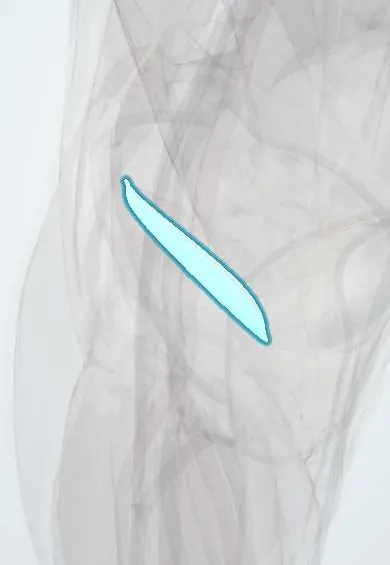
Lateral retinaculum !
The lateral retinaculum is a strong fibrous sheet on the outer side of the kneecap; it helps hold the kneecap in its groove.
Official name: Lateral patellar retinaculum
Opens in the Durgical app, no sign-up needed.
What it is
A fibrous sheet on the outer side of the kneecap.
What it does
Helps keep the kneecap centred; if too tight it can pull the kneecap outward.
Key facts
- Attaches
- Expansions of vastus lateralis and iliotibial tract: lateral patellar border to lateral tibial condyle
- Function
- Lateral patellar stabilizer; when tight it tilts the patella laterally
- Tight in
- Knee flexion; medial glide of patella
Relations
- Superficial
- IT band fibres (superficial oblique retinaculum) and skin
- Deep
- Joint capsule and synovium over the lateral femoral condyle and the lateral trochlea
- Above
- Vastus lateralis, whose aponeurosis forms part of it
- Below
- Lateral border of the patellar tendon and the lateral tibial condyle near Gerdy tubercle
How to find it
With the knee straight and the quadriceps relaxed, it is the firm sheet felt between the outer edge of the patella and the IT band; a tight one resists lifting the lateral edge of the patella and pushing the patella medially. Tenderness along the lateral facet goes with lateral overload.
Examination
- Patellar tilt test: knee straight, the lateral edge of the patella is lifted; failure to raise it to horizontal means a tight lateral retinaculum
- Medial patellar glide: movement of a quarter of the patellar width or less suggests tight lateral restraints
On imaging
Axial (skyline) X-ray, CT or MRI shows patellar tilt, lateral facet narrowing and, after a dislocation, the retinacular tear. A lateral release is visible on MRI as a gap in the retinaculum.
Clinical notes
- Lateral release can be combined with medial stabilization; on its own it does not treat lateral instability
- Iatrogenic medial patellar instability after an excessive release is a classic complication
Related conditions
Test yourself
Think of the answer, then tap the question to see it.
How does the lateral retinaculum compare with the medial retinaculum?
It is thicker and stronger, in two layers: a superficial oblique layer from the IT band and a deep transverse layer.
What does a tight lateral retinaculum cause?
It tilts the patella and overloads its lateral facet (excessive lateral pressure syndrome).
What is the classic complication of an excessive lateral release?
Iatrogenic medial patellar instability.
Educational information, not medical advice. For any symptom, see a health professional.
Its names
- Official Latin (TA2)
- retinaculum laterale patellae TA2 2624
- Official Spanish (DeCS)
- Retináculo lateral de la rótula
- English (TA2)
- Lateral patellar retinaculum
- Also called
- retinaculum fibulare patellae
Related
Sources: Terminologia Anatomica (TA2); Anatomy, Bony Pelvis and Lower Limb, Knee Patella; Anatomy of the Human Body (20th ed.): Surface Anatomy of the Lower Extremity (Lea & Febiger (Gray's Anatomy, 1918), 1918) · adapted, Public domain.
Spotted a mistake?We are always improving for the community. Tell us what is wrong and our team will review it.Report a mistake
Representative model, for educational purposes only.