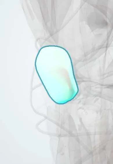
Patellar fracture !
A patellar fracture is a break of the kneecap; when the pieces separate, the knee may no longer be able to straighten.
Updated 2026-10-29
Structures involved
Tap one to see what it is and open it in 3D.
- Patella
- Patellar cartilage
- Medial retinaculum
- Lateral retinaculum
- Rectus femoris
- Vastus medialis
- Vastus lateralis
- Vastus intermedius
- Prepatellar bursa
- Joint capsule
In the clinical exam
What it is
A break of the kneecap (patella). Displaced fractures can disrupt the extensor mechanism and leave the patient unable to straighten the knee.
Who gets it
An uncommon fracture overall; most often seen in adults after a fall onto the knee or a dashboard impact in road accidents.
Causes
- Direct blow: fall onto the front of the knee, dashboard injury
- Indirect: violent quadriceps contraction against a flexing knee, causing a transverse fracture
Risk factors
- Falls in older adults
- Road traffic accidents
- Osteoporosis
- Previous knee surgery using the patella (e.g. some ligament reconstructions or knee replacement)
Pathophysiology
The patella is a sesamoid bone in the quadriceps tendon that increases the lever arm of the extensor mechanism. A transverse fracture with separation of the fragments, especially if the medial and lateral retinacula also tear, breaks the extensor mechanism so active knee extension is lost.
Classification
- Non-displaced or displaced (criteria vary; step-off and fragment gap)
- Pattern: transverse, vertical, comminuted (stellate), marginal, pole (sleeve in children)
- Open or closed
Symptoms and signs
- Pain and swelling at the front of the knee
- Haemarthrosis
- Inability to straighten the knee or lift the straight leg
- Palpable gap in the kneecap
- Bruising over the knee
Diagnosis
- Clinical
- Straight-leg raise and active extension test the extensor mechanism; a palpable gap supports displacement. Aspiration of haemarthrosis with local anaesthetic can help the exam.
- Tests
- Usually none; pre-operative tests if surgery is planned.
- Imaging
- AP, lateral and axial (sunrise) knee X-rays; the lateral shows displacement. CT for complex fractures. A bipartite patella (usually upper-outer corner, smooth edges, often both sides) can mimic a fracture.
Differential diagnosis
- Bipartite patella
- Quadriceps tendon rupture
- Patellar tendon rupture
- Patellar dislocation
- Tibial plateau fracture
- Prepatellar bursitis
Goal of treatment
Restore a smooth joint surface and an intact extensor mechanism so the patient can actively straighten the knee, with early motion to avoid stiffness.
Treatment
- Acute
- Initial splinting with the knee straight, analgesia and ice.
- Conservative
- Non-displaced fractures with an intact extensor mechanism: knee immobiliser or brace in extension, then progressive flexion.
- Surgical
- Displaced fractures or loss of active extension: open reduction and internal fixation (e.g. tension-band wiring or screws) with retinacular repair; partial patellectomy for severe comminution.
- Rehabilitation
- Physiotherapy for range of motion and quadriceps strengthening.
Complications
- Post-traumatic patellofemoral arthritis
- Knee stiffness and weak extension
- Symptomatic hardware needing removal
- Loss of fixation or non-union
- Infection
- Deep vein thrombosis
Prognosis
Most patients regain good function. Comminuted fractures and joint-surface step-offs raise the risk of anterior knee pain and arthritis.
Test yourself
Think of the answer, then tap the question to see it.
Which bedside test assesses the extensor mechanism in a suspected patellar fracture?
The straight-leg raise: the patient tries to lift the leg with the knee fully straight. Inability suggests extensor mechanism disruption.
Which X-ray view best shows displacement of a patellar fracture?
The lateral view of the knee.
What normal variant can be mistaken for a patellar fracture?
Bipartite patella: a smooth, rounded fragment usually at the upper-outer corner, often seen on both sides.
When is surgery usually indicated for a patellar fracture?
When the fracture is displaced, the joint surface has a step, or the patient cannot actively extend the knee.
Related conditions
- Patellar Dislocation
- Knee Osteoarthritis
- Deep vein thrombosis
- Tibial Plateau Fracture
- Prepatellar Bursitis
- Hip dislocation
Educational information, not medical advice. For any symptom, see a health professional.
Sources: Anatomy, Bony Pelvis and Lower Limb, Knee Patella (StatPearls (NCBI Bookshelf), 2023).
Spotted a mistake?We are always improving for the community. Tell us what is wrong and our team will review it.Report a mistake
Representative model, for educational purposes only.