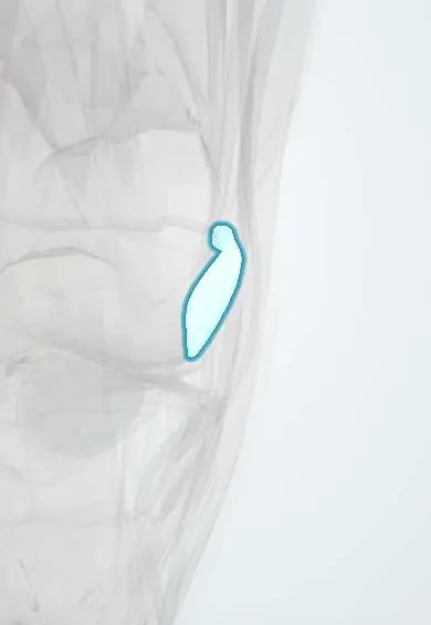
Medial retinaculum !
The medial retinaculum is a fibrous sheet on the inner side of the kneecap; it helps keep the kneecap centred in its groove.
Official name: Medial patellar retinaculum
Opens in the Durgical app, no sign-up needed.
What it is
A fibrous sheet on the inner side of the kneecap.
What it does
Helps keep the kneecap centred in its groove.
Key facts
- Attaches
- Vastus medialis aponeurosis expansion: medial patellar border and patellar ligament to medial tibial condyle; includes MPFL
- Function
- Resists lateral displacement of patella; reinforces anteromedial capsule
- Tight in
- Lateral glide of patella near full extension
Relations
- Superficial
- Deep fascia and skin; the infrapatellar branch of the saphenous nerve crosses below the patella
- Deep
- Joint capsule and synovium, with a medial plica in some knees, over the medial femoral condyle and trochlea
- Above
- Vastus medialis obliquus, whose aponeurosis blends into it
- Medial
- Superficial MCL and the adductor tubercle
How to find it
With the knee straight and the quadriceps relaxed, the patella can be moved sideways; the medial retinaculum is the sheet felt between its inner edge and the medial femoral epicondyle and adductor tubercle. After a lateral dislocation that whole line is often tender, mostly at the femoral end.
Examination
- Patellar apprehension test (Fairbank): knee at 20–30° of flexion, patella pushed laterally; fear or a quadriceps contraction to stop it means lateral instability
- Lateral patellar glide: the patella moving more than about half its width laterally, or a soft end point, suggests deficient medial restraints
On imaging
MRI after a dislocation shows tears or oedema of the medial retinaculum and MPFL, bone bruises on the medial patellar facet and the lateral femoral condyle, and any osteochondral fragment. Axial images show where the tear is.
Clinical notes
- Cutting the infrapatellar branch of the saphenous nerve during medial approaches leaves numbness on the front of the knee, lateral to the scar
- Medial plication tightens it, but on its own it has largely been replaced by MPFL reconstruction for recurrent instability
Related conditions
Test yourself
Think of the answer, then tap the question to see it.
Which ligament inside the medial retinaculum is the main restraint to lateral patellar displacement?
The medial patellofemoral ligament (MPFL); the patellomeniscal and patellotibial ligaments are secondary.
When is the medial retinaculum torn?
In almost every lateral patellar dislocation, usually together with the MPFL.
Which nerve can be cut in medial knee approaches through the retinaculum, and with what result?
The infrapatellar branch of the saphenous nerve, leaving numbness on the front of the knee, lateral to the scar.
Educational information, not medical advice. For any symptom, see a health professional.
Its names
- Official Latin (TA2)
- retinaculum mediale patellae TA2 2623
- Official Spanish (DeCS)
- Retináculo medial de la rótula
- English (TA2)
- Medial patellar retinaculum
- Also called
- retinaculum tibiale patellae
Related
Sources: Terminologia Anatomica (TA2); Anatomy, Bony Pelvis and Lower Limb: Medial Patellofemoral Ligament; Anatomy of the Human Body (20th ed.): Surface Anatomy of the Lower Extremity (Lea & Febiger (Gray's Anatomy, 1918), 1918) · adapted, Public domain.
Spotted a mistake?We are always improving for the community. Tell us what is wrong and our team will review it.Report a mistake
Representative model, for educational purposes only.