IT band !
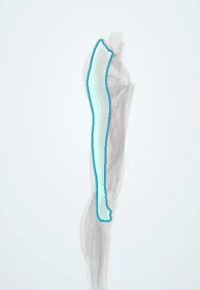
The iliotibial (IT) band is a long, tough strip running from the hip down the outer thigh to the tibia; it steadies the hip and knee.
Official name: Iliotibial tract
Opens in the Durgical app, no sign-up needed.
What it is
The iliotibial (IT) band: a long, tough band of fascia from the pelvis down the outer thigh to the outer tibia. The TFL and gluteus maximus pull on it from above.
What it does
Steadies the outer side of the hip and knee.
Key facts
- Attaches
- Thickening of fascia lata: iliac tubercle, receiving TFL and gluteus maximus, to Gerdy tubercle of lateral tibial condyle
- Function
- Transmits pull of TFL and gluteus maximus; stabilizes lateral hip and extended knee in stance
- Tight in
- Hip extension and adduction (Ober test position)
Relations
- Course
- From the iliac tubercle of the iliac crest, receiving TFL in front and much of gluteus maximus behind, down the lateral thigh to Gerdy tubercle, with fibres to the lateral patella
- Deep
- Vastus lateralis and the lateral intermuscular septum; at the knee, the lateral femoral epicondyle, with fat or a bursa between
- Behind
- Biceps femoris; at the knee its tendon lies behind the lower end of the band, with the LCL between
How to find it
Standing with the knee straight and weight on the leg, it is felt as a firm ridge on the outer thigh just above the knee, in front of the biceps tendon, ending on Gerdy tubercle lateral to the tibial tuberosity.
Examination
- Ober test: lying on the other side, the upper hip is extended and abducted and then released; if the thigh stays up instead of dropping, the band and TFL are tight
- Noble compression test: pressure over the lateral epicondyle while the knee is extended from 90°; pain at about 30° suggests IT band syndrome
On imaging
Rarely needed. In IT band syndrome, MRI shows fluid or oedema deep to the band over the lateral femoral epicondyle; ultrasound may show a thickened band.
Common problems
- IT band syndrome: pain on the outer knee, common in runners and cyclists.
- Outer snapping hip: the band catches over the greater trochanter with a snap on the outer hip.
How it is diagnosed
Physical exam; imaging is rarely needed.
Treatment
- Rest from the painful activity, stretching and hip strengthening, changes to training; injections occasionally. Surgery is rarely needed.
Clinical notes
- A strip of the band is used in lateral extra-articular tenodesis, added to ACL reconstruction to control rotation
- Lateral approaches to the femur and hip split the band in line with its fibres
- A Segond fracture avulses the lateral tibial rim at the attachment of the anterolateral structures, close to Gerdy tubercle
Related conditions
Test yourself
Think of the answer, then tap the question to see it.
Where and at what knee angle does IT band syndrome cause pain?
Over the lateral femoral epicondyle at about 30° of knee flexion, where the iliotibial band slides across it.
Where does the iliotibial (IT) band insert distally?
On Gerdy tubercle of the lateral tibial condyle, lateral to the tibial tuberosity.
What does a positive Ober test show?
Tight IT band and TFL: lying on the other side, the extended, abducted upper thigh stays up instead of dropping when released.
How does the IT band explain the pivot shift?
Its pull lies in front of the knee axis in extension and behind it beyond about 30° of flexion, when the subluxed tibia snaps back.
Educational information, not medical advice. For any symptom, see a health professional.
Its names
- Official Latin (TA2)
- tractus iliotibialis TA2 2690
- Official Spanish (DeCS)
- Tracto iliotibial DeCS D058745
- English (TA2)
- Iliotibial tract
- Also called
- iliotibial band · bandelette of Maissiat · tractus Maissiati
Related
Sources: Terminologia Anatomica (TA2); DeCS; Anatomy, Bony Pelvis and Lower Limb, Iliotibial Band (Tract).
Spotted a mistake?We are always improving for the community. Tell us what is wrong and our team will review it.Report a mistake
Representative model, for educational purposes only.